
By EIV Diagnostics · August 18, 2026
AI in Pathology: What Clinicians Need to Know
Discover how AI in pathology enhances decision-making and workflow, supporting clinicians with faster analysis and improved accuracy.
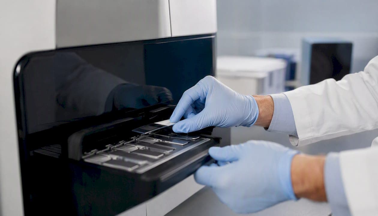
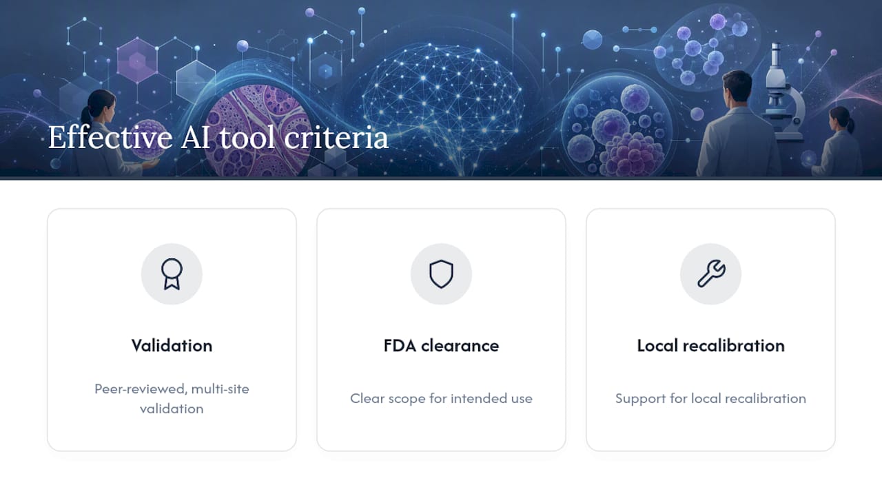
AI in pathology functions today as clinical decision support and workflow automation, not a replacement for the pathologist’s judgment. The technology handles narrowly defined, high-signal tasks: flagging suspicious regions, counting mitoses, quantifying biomarkers, and running quality checks on scanned slides before a human ever renders a diagnosis. That distinction matters for how you evaluate any AI claim: look for peer-reviewed validation at multiple sites, ask whether the tool carries an FDA clearance or qualification for its specific use, and confirm the vendor supports local recalibration rather than a one-size-fits-all model.
What changes in practice right now:
- Triage and QC tasks (focus detection, tissue folding, slide adequacy) run faster and more consistently than manual spot checks.
- Detection and counting tasks (lymph node metastases, mitotic figures, tumor cellularity) act as a second reader, catching outliers before sign-out.
Key Takeaways
AI in pathology works best as a second reader for narrowly defined tasks, and it only earns clinical trust through local validation, not vendor benchmarks alone.
| Point | Details |
|---|---|
| AI augments, doesn’t replace | Pathologists remain the final decision-maker; AI speeds triage and standardizes quantification. |
| Match evidence to the claim | Prioritize FDA-cleared tools and prospective reader-assistance studies over retrospective benchmarks alone. |
| Domain shift is the top risk | Revalidate locally after any scanner, stain, or software change using a fixed challenge set. |
| Build guardrails into the UI | Require an independent pathologist read before showing AI output to limit automation bias. |
| EIV Diagnostics supports validation | Its molecular, digital, and histopathology services can serve as a benchmarking partner for labs piloting AI tools. |
Table of Contents
- How Is AI Used in Pathology Today?
- What Evidence Supports AI Tools in Pathology?
- How Does AI Fit Into the Digital Pathology Workflow?
- What Are the Biggest Risks With AI in Pathology?
- What Should a Lab Do Before Using AI Clinically?
- What Deployment Models Exist for Pathology AI?
- Where EIV Diagnostics Fits Into Your AI Adoption Plan
- Sources
- FAQ
How Is AI Used in Pathology Today?
The strongest evidence clusters around a handful of well-defined tasks, while broader multimodal applications remain earlier stage. Here’s where AI in pathology currently earns its place in the workflow:
- Screening and triage — Pre-sorting slides by likelihood of malignancy so pathologists review high-risk cases first. Evidence tier: retrospective validation, moving into prospective workflow trials.
- Lesion and metastasis detection — Flagging lymph node micrometastases or small foci easy to miss on a first pass. Evidence tier: reader-assistance studies showing reduced miss rates.
- Mitosis counting and tumor cellularity — Automating a notoriously tedious, high-variability manual count. Evidence tier: strong retrospective and some prospective data.
- IHC and biomarker scoring — Quantifying HER2, Ki-67, and PD-L1 staining with reproducible thresholds instead of eyeballed percentages. Evidence tier: regulatory-cleared tools exist for several markers.
- Prognostic and biomarker prediction — Correlating morphology with likely outcomes or treatment response. Evidence tier: mostly research-stage, promising but not routine.
- Quality control — Catching scanning artifacts, poor focus, or folded tissue before a case reaches a pathologist. Evidence tier: mature, widely deployed.
- Research and drug development — Quantifying tumor microenvironment features across large cohorts for pharma studies. Evidence tier: research use, not diagnostic.
Foundation models that combine image data with molecular and clinical inputs are an active research direction. They’re worth watching, but multimodal diagnostic tools are not yet at the clinical maturity of single-task detection or scoring algorithms, a gap the 2026 review of digital and computational pathology advances lays out in detail.
What Evidence Supports AI Tools in Pathology?
Not all AI claims carry equal weight, and the evidence tier behind a tool tells you how much to trust it. Retrospective, single-site validation is the weakest signal. Multi-site retrospective studies are better. Prospective reader-assistance trials, where pathologists use the tool live and results get compared to unassisted reads, carry real clinical weight. Regulatory clearance, particularly an FDA 510(k) clearance or qualification for a specific intended use, is the strongest available signal, though it still says nothing about performance in your specific lab’s population and scanner setup.
- Peer-reviewed multi-site validation studies remain the baseline for trusting a sensitivity or specificity claim.
- FDA 510(k) clearances exist for select applications like prostate cancer detection and IHC quantification, but clearance scope is narrow and use-case specific.
- Documented hospital pilot deployments show what happens outside a controlled study.
Statistic Callout: A Stanford Medicine pilot of the AI tool Nuclei.io found that pathologists using it worked faster and with improved diagnostic accuracy in real clinical settings, a rare example of prospective, in-workflow performance data rather than a retrospective benchmark. That gap between retrospective benchmarks and real-world workflow data is exactly why systematic reviews note more heterogeneity in study design for prospective deployment evidence than for algorithm accuracy claims.
How Does AI Fit Into the Digital Pathology Workflow?
AI touches several discrete points between slide preparation and sign-out, and each one carries its own integration burden.
- Pre-analytic standardization. Fixation time, staining protocol, and section thickness all shift how an image looks to a model, long before scanning starts.
- Whole-slide imaging. The scanner and its settings become part of the model’s input distribution. Switching scanner brands without revalidating is a common cause of silent accuracy drops.
- Model inference and QC. The algorithm runs, flags regions or generates scores, and a QC layer checks for out-of-distribution inputs.
- Human-in-the-loop review. A pathologist reviews AI output alongside the slide, not instead of it, and renders the final diagnosis.
- Audit logs and monitoring. Every AI-assisted case gets logged for later concordance review and drift detection.
Integration depends on whether your LIS/PACS can accept structured AI outputs, how much latency your workflow tolerates, and whether cloud inference fits your data governance policy or you need an on-premises deployment. Labs weighing these tradeoffs often start by reviewing how existing digital pathology providers structure their integration models before committing to one.
Pro Tip: Keep a fixed local “challenge set” of representative slides and rerun it after any scanner firmware update, staining protocol change, or model version upgrade. Silent domain shift is far easier to catch against a known baseline than to discover after a diagnostic miss.
What Are the Biggest Risks With AI in Pathology?
The most common real-world failure isn’t a bad algorithm. It’s a good algorithm meeting a scanner, stain batch, or patient population it wasn’t trained on. This domain shift problem explains most of the gap between published accuracy and routine performance.
- Domain and stain shift — A model trained on one scanner or stain vendor can quietly underperform on another.
- Shortcut learning — Models sometimes key on artifacts (ink marks, slide labels) rather than genuine pathology.
- Miscalibrated probabilities — A confidence score of 90% doesn’t always mean 90% accuracy; calibration needs its own validation.
- Misleading overlays — Heatmaps and explanation visuals can look convincing while pointing at the wrong tissue feature.
- Biased or leaked training data — Overlap between training and test sets, or underrepresented subgroups, inflates reported performance.
Guard against these with local calibration before go-live, external validation across sites and time periods, uncertainty quantification rather than flat confidence scores, and a concordance log that tracks every disagreement between the AI and the final pathologist call.
Pro Tip: Build a UI guardrail that requires the pathologist to form an independent impression before the AI output displays, or reveal AI findings progressively rather than all at once. This single design choice reduces automation bias more reliably than any disclaimer text.
What Should a Lab Do Before Using AI Clinically?
- Define the intended use and claim. Know exactly what the tool is validated to do, not what it might do.
- Assemble a representative local dataset. Include your own scanners, stains, and patient mix.
- Run retrospective external validation. Confirm performance holds outside the vendor’s original study population.
- Pilot as a reader-assistance study. Compare AI-assisted reads to unassisted reads in your own workflow.
- Complete regulatory and QA documentation. Match the tool’s clearance scope to your actual use case.
- Integrate with LIS/PACS and set up monitoring. Build the audit trail before, not after, go-live.
- Train staff and establish governance. Assign clear ownership for ongoing oversight.
Rough timelines: a reader-assistance pilot typically runs 3 to 6 months; full prospective validation across sites often takes 6 to 12 months. Success needs a pathologist lead, a lab information manager, a data scientist or IT partner, and someone owning vendor procurement and contract terms.
What Deployment Models Exist for Pathology AI?
Labs adopt AI through a few recognizable patterns, each suited to a different scale and risk tolerance.
- Cloud inference fits labs that want to avoid managing GPU infrastructure and can accept data leaving the building under a business associate agreement.
- On-premises appliances suit labs with strict data residency requirements or high case volumes where latency matters.
- Integrated LIS/PACS plugins slot AI output directly into the existing sign-out screen, minimizing workflow disruption.
- Federated learning collaborations let multiple institutions train a shared model without pooling raw patient data.
- Bespoke local algorithms serve niche research questions where no commercial tool exists yet.
Most tools move from research prototype to task-specific validation to institutional integration, with evidence strength increasing at each step. Enterprise integrated platforms tend to carry the most validation; entry-level field apps and population-scale screening services trade some rigor for speed and reach.
A pragmatic view from EIV Diagnostics
We treat AI as a tool to speed turnaround and standardize quantitative readouts, never as a substitute for pathologist judgment. That means local validation, documented QA, and board-certified oversight before any AI-assisted result reaches a report.
Where EIV Diagnostics Fits Into Your AI Adoption Plan
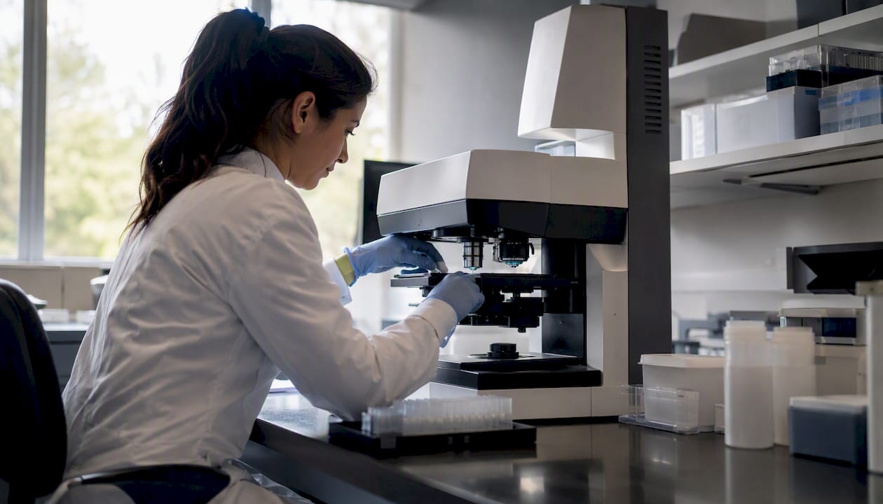
Piloting AI in pathology means validating it against real specimens, real turnaround pressure, and real reporting standards, not a vendor’s marketing deck. EIV Diagnostics runs molecular pathology services, digital pathology workflows, and histopathology processing built around board-certified pathologist oversight and rapid, precise reporting, which makes it a practical partner when your lab needs a local validation dataset or a second-opinion workflow to benchmark a new algorithm against.

For labs and providers assembling a representative case set for external validation, EIV Diagnostics also offers mobile phlebotomy to simplify specimen logistics without adding a patient visit to an already busy clinic schedule. If you’re planning a pilot, a validation partnership, or simply need a second-read baseline while you calibrate a new tool, reach out through EIV Diagnostics to start the conversation.
Sources
- Artificial intelligence in digital pathology diagnosis and analysis: technologies, challenges, and future prospects - PMC
- What’s new in digital and computational pathology 2026: advances in adoption, standards, AI technologies, and clinical integration
- AI tool gives pathologists speed, accuracy and a new way to …
This article is general information, not a substitute for advice from a qualified doctor. Consult a qualified healthcare professional about your own circumstances before acting on anything here.
FAQ
Is Pathology Going to Be Replaced by AI?
No. Current evidence and clinical practice position AI as decision support that speeds tasks like counting and quantification, while final diagnosis and clinical judgment stay with the pathologist.
How Is AI Used in Pathology Right Now?
AI supports screening and triage, mitosis counting, biomarker scoring, lesion detection, and slide quality control, with the strongest evidence behind narrowly defined, high-signal tasks.
What Makes an AI Pathology Tool Effective?
Effective tools carry peer-reviewed multi-site validation, a clear FDA clearance scope for their intended use, and support for local recalibration; labs like EIV Diagnostics evaluate tools against these criteria before trusting them in a workflow.
What Diseases Can AI Help Diagnose in Pathology?
AI-assisted tools show the most validated impact in cancer pathology, including breast, prostate, and lymph node metastasis detection, along with biomarker scoring for markers like HER2 and PD-L1.
Recommended
- Clinical Pathology Labs: What Patients Need to Know | EIV Diagnostics | EIV Diagnostics
- Clinical Pathology Services | EIV Diagnostics | EIV Diagnostics | EIV Diagnostics
- Molecular Diagnostic Testing: What Patients and Clinicians Need to Know | EIV Diagnostics | EIV Diagnostics
- Dermatopathology Services: What Patients and Providers Need to Know | EIV Diagnostics | EIV Diagnostics