
By EIV Diagnostics · October 25, 2026
Urine Microscopic Exam: What It Shows and How to Read Results
A urine microscopic exam looks at cells, casts, crystals and bacteria in your sample. Learn how to prepare, what results mean, and when cytology is needed.
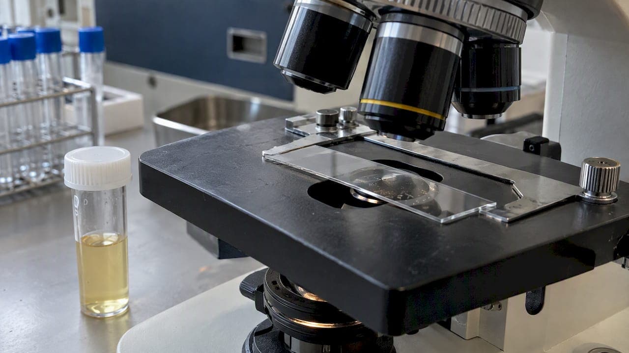
A urine microscopic exam is the part of a urinalysis where a technologist or pathologist looks at your urine under a microscope. It finds things a dipstick cannot see: red and white blood cells, casts, crystals, bacteria, yeast and abnormal cells shed from the urinary tract. MedlinePlus describes urinalysis as a physical, chemical and microscopic examination of urine, and the microscopic step is the one that confirms what the other two only hint at.
This guide explains what happens to your sample, how to collect it so the result is trustworthy, what the numbers on your report mean, and when a different test, such as urine cytology, is the right next step.
TL;DR
- A urine microscopic exam examines concentrated urine sediment for cells, casts, crystals and microorganisms. It often follows an abnormal dipstick, but it can also be ordered on its own.
- Preparation matters: a clean-catch, midstream sample, no hard exercise beforehand, and no collection during your period. A poorly collected sample is the most common reason for a confusing result.
- One abnormal result rarely means a diagnosis. Doctors usually repeat the test before drawing conclusions.
- Urine cytology is a different test. It looks for abnormal cells, mainly to evaluate or follow bladder cancer, and it does not replace routine microscopy.
Table of Contents
- What a urine microscopic exam actually is
- What the microscope looks for
- How to collect a sample that gives a reliable result
- Reading your results without panicking
- Urine microscopy versus urine cytology
- What we see from the lab side
- Getting a urine test in the Central Valley
- Questions to ask before and after your test
- Frequently asked questions
- Sources
What a urine microscopic exam actually is
The test is one of three parts of a complete urinalysis. StatPearls is not our source here, so we will lean on the clinical references we checked: the physical exam looks at color and clarity, the chemical exam uses a dipstick to check for things like protein, glucose, blood and leukocyte esterase, and the microscopic exam looks at the solid material in the urine.
In the standard method, the lab spins the urine in a centrifuge so that cells and particles collect at the bottom. A 2024 review of urine microscopy in kidney disease describes the typical protocol: about 10 mL of urine spun at roughly 400g for 5 minutes, the liquid on top removed, and the pellet resuspended in the small amount left behind. A drop goes on a slide and is examined at low and high power.

The result is reported as counts per field, for example red blood cells per high-power field (RBC/HPF), plus descriptions such as “few bacteria” or “hyaline casts present.”
Why the dipstick is not enough
A dipstick is a fast screen. It detects chemical reactions, not what is physically in the urine. A dipstick can say there is blood, but it cannot tell you whether the red cells look like they came from the kidney’s filters or from a bladder irritation. It also can’t identify casts or crystals. The microscopic exam adds that detail, and for that reason clinicians often order it when the dipstick is positive for blood, protein, nitrite or leukocyte esterase.
What the microscope looks for
Here is what each finding generally points to. None of these is a diagnosis on its own, and your clinician reads them alongside your symptoms, history and other labs.
| Finding | What it is | What it may suggest |
|---|---|---|
| Red blood cells | Blood cells in the urine (hematuria) | Infection, kidney stones, kidney filter disease, bladder or kidney tumors, or a contaminated sample |
| White blood cells | Infection-fighting cells (pyuria) | Urinary tract infection, inflammation, kidney infection |
| Epithelial cells | Cells lining the urinary tract or skin | Few is normal; many suggest the sample was contaminated |
| Casts | Protein molds formed in kidney tubules | Type matters: hyaline casts can be harmless, cellular or granular casts point to kidney disease |
| Crystals | Mineral deposits | Often harmless, sometimes linked to stones or metabolic problems |
| Bacteria, yeast, parasites | Microorganisms | Infection, or contamination during collection |
MedlinePlus lists urinary tract infections, glomerulonephritis, kidney stones, bladder cancer and diabetes-related kidney damage among the conditions urinalysis can help detect or monitor. It also gives a general reference range: red and white cells should be near 0 to 5 per high-power field, though labs set their own ranges.
Red blood cells and the 3-per-field threshold
Blood you cannot see is called microscopic hematuria. The American Urological Association defines it as three or more red blood cells per high-power field on microscopy, and its risk-based approach looks at your age, smoking history and how many cells were counted to decide how far to investigate. The same summary notes that a single urinalysis is enough to detect about 95% of microscopic hematuria, so a positive result is a reason to talk to your clinician, not to repeat tests on your own.
Casts and dysmorphic cells
Casts and misshapen red cells are where manual review earns its keep. The kidney review cited above notes that automated analyzers are fast and standardized but can miss non-hyaline casts, dysmorphic red blood cells and certain crystals that an experienced reader can spot under the microscope. Those findings help separate kidney filter disease such as glomerulonephritis from bleeding further down the urinary tract. When a result is unclear, ask whether a person reviewed the slide.
How to collect a sample that gives a reliable result
Most “abnormal” microscopic results in healthy people trace back to the sample, not the patient. Follow these steps.
- Check the timing. Avoid heavy exercise, a very high-protein meal and sexual activity for about 24 hours before collecting. A 2024 review of diet, drugs and exercise effects on urine reports these can add red cells, epithelial cells, protein or sperm to the sample.
- Skip menstruation if you can. The same review advises against collecting during your period or in the few days around it, because menstrual blood can be mistaken for hematuria. If your test cannot wait, tell the lab or your clinician so the result is read in context.
- Wash first. InformedHealth.org advises cleaning the genital area with water, no soap, so bacteria and other substances do not contaminate the urine.
- Use the midstream method. Begin urinating into the toilet, then catch the middle of the stream in the cup. Discarding the first few seconds reduces skin bacteria in the sample.
- Close and label the cup. Hand it in promptly.
Tell whoever orders the test about supplements, medicines and recent illness. Some drugs and vitamins change urine color and chemistry, and your clinician needs to know when interpreting the report.
Why speed matters after collection
Urine changes quickly once it leaves the body. The kidney review says analysis should ideally occur within one hour of voiding at room temperature. Waiting too long lets cells break down, pH shift and bacteria multiply, which alters the sediment. Refrigeration slows bacteria but can cause phosphates and urates to precipitate as crystals that look like a finding but are not. If you collect at home, ask the lab in advance how to store and deliver the sample. That is one reason some patients prefer to have a trained phlebotomist handle the logistics.
Reading your results without panicking
A report with a flagged value is stressful. A few principles help.
One result is a data point. The National Kidney Foundation’s guidance on albuminuria and proteinuria explains that confirming chronic protein in the urine takes repeated tests over months, in part because exercise, dehydration and diet can cause temporary changes. The same logic applies to microscopic findings: a single borderline count is usually followed by a repeat.
Contamination looks like disease. A primary care review from LSU notes that more than 10 epithelial cells per high-power field generally suggests a contaminated sample. If your report mentions many squamous cells with bacteria and white cells, the lab may recommend a recollection before anyone treats an infection.
Context decides everything. Red cells with burning and urgency point one way. Red cells with swelling, high blood pressure or foamy urine point another. The National Kidney Foundation notes that lots of persistent foam can signal protein in the urine, one of the early signs the kidneys are not working as they should.

When to call your clinician promptly
Contact a doctor soon if you can see blood in your urine, if you have fever with back or flank pain, if you cannot urinate, or if you are pregnant and your report shows infection. Visible blood always deserves an evaluation, even if it comes and goes. If you have severe pain, are confused, or feel faint, go to urgent care or call 911.
Urine microscopy versus urine cytology
Readers often confuse these tests, and search results mix them. They answer different questions.
| Urine microscopic exam | Urine cytology | |
|---|---|---|
| Main question | What cells, casts, crystals and microbes are in the urine? | Do any cells look cancerous or precancerous? |
| Who reads it | Lab technologist, sometimes a pathologist or nephrologist | Cytotechnologist and pathologist |
| Typical use | Infection, kidney disease, stones, unexplained hematuria | Evaluating blood in the urine for urothelial cancer, and following people treated for bladder cancer |
| Sample | Fresh midstream urine | Fresh urine, handled to preserve cell detail |
Cytology is more specialized. The cells are stained and examined for changes in size, shape and nuclear features. It tends to catch higher-grade tumors better than low-grade ones. A published comparison of cytology and a FISH assay in 1,835 urine samples described cytology as limited by low sensitivity and found that the FISH test detected more urothelial cancers but also produced more false positives. That is why a negative cytology does not close the case when your symptoms or risk factors say otherwise, and why an atypical result is often followed by cystoscopy or another test.
If you are asking about urine cytology for hematuria, the practical answer is this: your clinician decides whether it is needed based on your age, smoking history, the amount of blood, and your symptoms. It is not a routine add-on for every positive dipstick.
What we see from the lab side
We are a pathology laboratory, so we spend our days on the step between a collected specimen and a report a clinician can act on. A few points shape how we think about urine testing.
Technique drives reliability. The kidney microscopy review points out that non-standardized methods can vary enormously, with coefficients of variation reaching 100%. The spin speed, the volume, the way the pellet is resuspended and who looks at the slide all change what a patient is told. A reading is only as good as the process behind it.
Automation is useful, not complete. Analyzers are the right tool for high volumes and for clearing normal samples. When an analyzer flags a sample, or when the clinical picture does not fit the numbers, a human review of the sediment adds information. Board-certified pathologists exist for exactly those judgment calls.
The sample is half the test. Many of the problems we would prevent happen before the specimen arrives: the wrong container, a delay, a warm car, a contaminated catch. That is part of why we offer mobile phlebotomy, so specimen collection and handling are done by trained staff rather than left to chance.
Patients deserve plain-language results. A report full of abbreviations helps no one. Whatever test you take, you should be able to ask your clinician or us what each flag means and what happens next.
— EIV Diagnostics
Getting a urine test in the Central Valley
EIV Diagnostics is an independent pathology laboratory in Fresno, serving patients and providers across Fresno, Clovis, Madera, Tulare, Kings and the surrounding Central Valley communities. Our clinical pathology services and cytology work are overseen by board-certified pathologists, and the lab holds CAP accreditation.
You can reach us in a few ways:
- With a doctor’s order: bring your requisition, or ask your provider to send it to us.
- Without an order: our self-pay testing option lets you purchase lab tests directly if you want to check your health without a doctor’s visit first. Self-pay is a good way to get a baseline, but an abnormal result still needs a clinician to interpret it.
- At home or at work: mobile phlebotomy starts from $65 per visit, and a trained phlebotomist comes to you.
Browse the full list of what we offer on our services page, including cytology, histopathology and toxicology. If you are not sure which test fits your situation, contact us before ordering. We can tell you what we offer, and your own clinician can tell you what you need.
Questions to ask before and after your test
Going in prepared makes the result more useful.
- Is this a dipstick only, or does it include microscopy?
- Will a person review the slide if the analyzer flags something?
- Do I need to fast, or can I eat and drink normally? Staying hydrated helps most people produce a usable sample, and you should follow your provider’s instructions.
- If a value is abnormal, will the test be repeated and when?
- If there is blood, what is the plan to find the source, and is cytology part of it?
Frequently asked questions
What diseases can a urine microscopic exam help diagnose?
It can help detect or monitor urinary tract infections, kidney stones, glomerulonephritis, diabetes-related kidney damage and bladder cancer, according to MedlinePlus. It does not diagnose any of them alone. The findings point your clinician toward the next test.
What should I avoid before giving a urine sample?
Avoid hard exercise, a very high-protein meal and sexual activity for about 24 hours, and do not collect during your period if you can avoid it. Research on diet and exercise effects shows these can add cells or protein that mimic disease. Use a clean-catch midstream sample and wash with water only.
Can dehydration cause blood in the urine?
Dehydration and strenuous exercise can cause temporary changes in urine, and the National Kidney Foundation notes that such factors often explain brief variations. That does not mean you can assume it is harmless. If blood shows up on a test, your clinician should decide whether to repeat it or investigate further, and you should see a doctor right away for visible blood.
Does a normal urine microscopic exam rule out bladder cancer?
No. Microscopy checks for red cells and other elements but does not examine cells for cancerous features. Cytology and cystoscopy are used for that purpose, and your clinician chooses based on your risk. If you have visible blood or other symptoms, keep your appointment even if one result is normal.
Sources
- Urinalysis: MedlinePlus Medical Encyclopedia
- Diagnostic Utility of Urine Microscopy in Kidney Diseases
- Effects of Diet, Supplements, Drugs and Exercise on Urine Characteristics
- Understanding urine tests: InformedHealth.org
- Microscopic Hematuria in Adults: AUA Recommendations, American Family Physician
- Kidney Disease and Albuminuria/Proteinuria, National Kidney Foundation
- Signs and symptoms of kidney disease, National Kidney Foundation
- Evaluation of UroVysion and cytology for bladder cancer detection, Cancer Cytopathology