
By EIV Diagnostics · August 16, 2026
Skin Cancer Diagnosis: What to Expect and Next Steps
Learn what to expect during a skin cancer diagnosis, from the clinical exam to biopsy and pathology results, guiding your next steps.
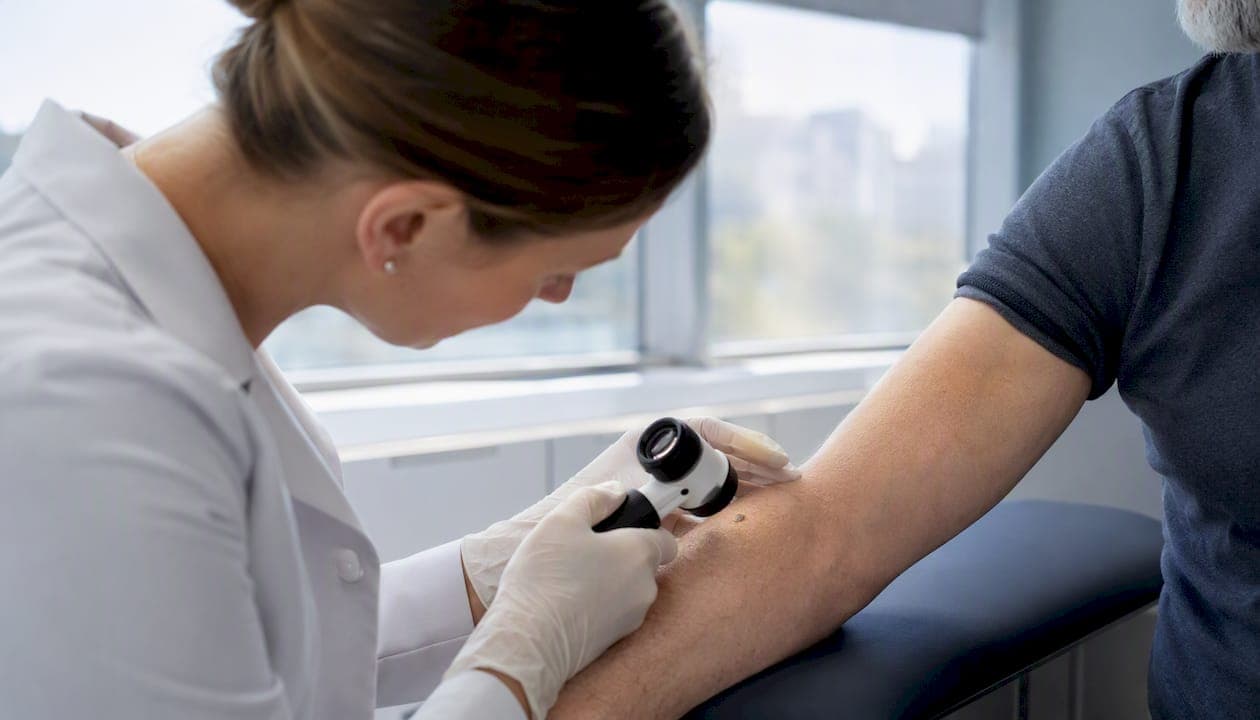
Skin cancer is diagnosed through a clinical skin exam followed by a tissue biopsy; the resulting pathology report then determines staging and drives every treatment decision that follows. If your provider spots a suspicious lesion, here is the sequence you will most likely move through:
- Clinical exam: Full-body visual inspection, often with dermoscopy, to identify lesions worth sampling
- Biopsy: A small tissue sample removed under local anesthesia, usually in under 30 minutes
- Pathology processing: Tissue is prepared, stained, and reviewed by a pathologist, typically within one to two weeks for routine cases
- Follow-up appointment: Your provider reviews the report, explains the findings, and outlines next steps
Guideline bodies including the American Academy of Dermatology (AAD), the National Comprehensive Cancer Network (NCCN), and the American Joint Committee on Cancer (AJCC) set the standards that govern how each step is performed and reported. When a dermatopathologist or a specialized lab such as EIV Diagnostics reviews the specimen, the report carries the precision those standards require.
Key Takeaways
Skin cancer diagnosis follows a defined sequence: clinical exam plus biopsy produces a pathology report that determines staging, which then drives every treatment and referral decision.
| Point | Details |
|---|---|
| Biopsy is required | No skin cancer diagnosis is definitive without tissue; excisional biopsy is preferred for suspected melanoma. |
| Pathology report elements matter | Breslow depth, ulceration, mitotic rate, and margin status are required for accurate AJCC staging. |
| Imaging is stage-dependent | Ultrasound, CT, PET, and MRI are used selectively for intermediate and advanced melanoma, not early-stage disease. |
| Molecular testing guides advanced treatment | BRAF and PD-L1 testing on biopsy tissue determines eligibility for targeted therapy and immunotherapy. |
| EIV Diagnostics | Offers board-certified dermatopathology, molecular pathology, and digital pathology review for patients and providers in the Central Valley. |
Table of Contents
- How skin cancer diagnosis begins: self-checks and the clinical exam
- In-office tools that help clinicians decide whether to biopsy
- Biopsy types: what each involves and what to expect
- Reading the pathology report: the terms that determine your stage
- When additional tests and imaging are recommended
- Molecular and genetic testing on biopsy tissue
- How long the diagnostic process usually takes
- What a confirmed diagnosis usually leads to next
- Signs that need faster evaluation
- How specialized diagnostic labs support accurate, timely results
- Why precision in diagnosis matters more than speed alone
- Advanced diagnostic services through EIV Diagnostics
- Sources
- FAQ
How skin cancer diagnosis begins: self-checks and the clinical exam
Patient self-exam and clinician skin exam are the most common entry points into the diagnostic process, though high-quality evidence that population-level screening reduces skin cancer deaths remains limited. What self-checks reliably do is get people into a clinic sooner.
What to look for on your own skin
The ABCDE framework is the standard self-exam guide:
- A — Asymmetry: One half of the spot does not match the other
- B — Border: Ragged, notched, or blurred edges
- C — Color: Multiple shades of brown, black, red, white, or blue within one lesion
- D — Diameter: Larger than 6 mm (about the size of a pencil eraser), though melanomas can be smaller
- E — Evolving: Any change in size, shape, color, or a new symptom such as bleeding or itching
Spots that bleed without trauma, crust repeatedly, or simply look different from everything else on your skin (“ugly duckling” sign) also warrant evaluation. Total-body photography and mole mapping are used in patients with many atypical nevi or a personal history of melanoma; sequential photos make subtle changes visible that the naked eye misses on a single visit.
What happens during a clinician’s exam
A dermatologist or primary care provider takes a focused history (how long the lesion has been there, any changes, personal or family history of skin cancer), then inspects the entire skin surface, not just the spot you came in about. Palpation of nearby lymph nodes follows when a lesion looks suspicious. Clinical photography is often taken to document baseline appearance.
Primary care typically refers to dermatology when a lesion has atypical features, sits in a cosmetically or functionally sensitive location, or when the patient has a prior skin cancer history. Uncertainty alone is a valid reason for referral.
Pro Tip: Photograph the lesion with your phone before your appointment, note the date you first noticed it, and write down any changes you have observed. Providers can compare your photos against the current appearance, which often shortens the time to a biopsy decision.
In-office tools that help clinicians decide whether to biopsy
Dermoscopy is the most widely used noninvasive tool for triaging skin lesions, and its impact on accuracy is well documented. According to the AAD, dermoscopy improves diagnostic accuracy and a dermatopathologist’s review is often used to confirm the diagnosis and subtype. Pooled data show dermoscopy raises melanoma sensitivity from roughly 76% to 92% and specificity from 75% to 95% compared with naked-eye inspection alone.
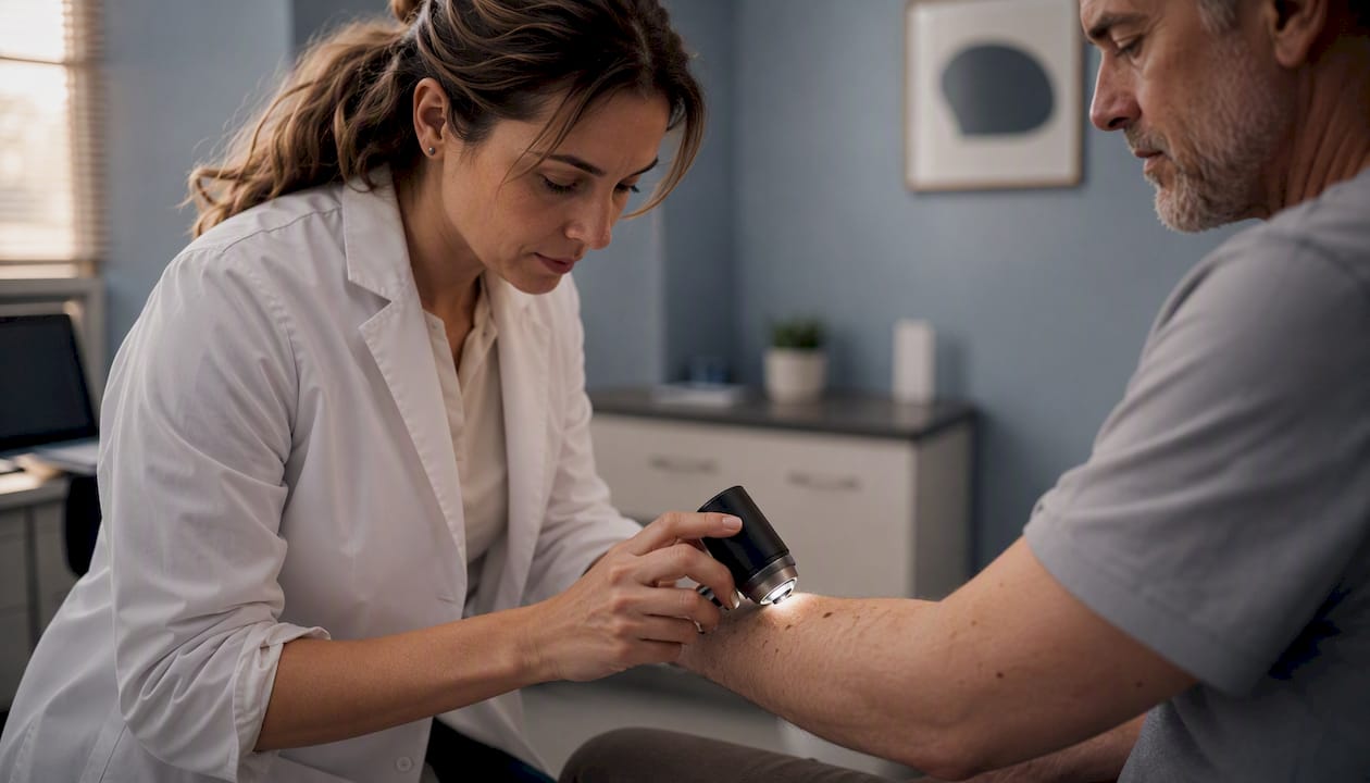
That improvement translates directly into fewer unnecessary biopsies and fewer missed cancers. Most dermatologists use a handheld dermoscope routinely; many primary care offices now have one as well.
Advanced tools available in specialty settings
Noninvasive imaging tools such as reflectance confocal microscopy (RCM), optical coherence tomography (OCT), and high-frequency ultrasound can reduce unnecessary biopsies in specialized settings, but they are typically available only in dermatology specialty or academic centers.
- Reflectance confocal microscopy (RCM): Provides near-histologic resolution of the epidermis and superficial dermis in real time; useful for ambiguous facial lesions where biopsy scarring is a concern
- Optical coherence tomography (OCT): Cross-sectional imaging of the skin to roughly 1–2 mm depth; used mainly for basal cell carcinoma assessment
- High-frequency ultrasound (20–75 MHz): Measures tumor thickness before excision and can assess lymph node architecture; more commonly used in Europe than in the United States
None of these tools replace biopsy. They inform the decision about whether and how to biopsy, and they help plan the procedure.
Biopsy types: what each involves and what to expect
A skin biopsy is required for definitive skin cancer diagnosis; excisional biopsy with narrow margins is preferred for suspected melanoma so Breslow depth can be measured accurately. The choice of technique depends on lesion size, location, and clinical suspicion.
| Biopsy Type | Typical Use Case | Tissue Depth | Cosmetic Consideration | Recommended When |
|---|---|---|---|---|
| Shave | Low-suspicion lesions, seborrheic keratoses, superficial BCCs | Epidermis to superficial dermis | Minimal scarring | Melanoma NOT suspected |
| Punch | Small lesions, inflammatory conditions, nail unit | Full thickness, small core | Small circular scar | Moderate suspicion; small lesion |
| Excisional | Suspected melanoma, small pigmented lesions | Full thickness with margins | Linear scar, may need closure | Melanoma suspected; lesion < 2 cm |
| Incisional | Large lesions where full removal is impractical | Partial full-thickness sample | Variable | Large acral or facial lesions |
What to expect during the procedure
Local anesthetic (typically lidocaine) is injected around the lesion. Most patients feel a brief sting from the injection, then nothing during the removal itself. The tissue is placed in formalin and sent to the pathology lab with a requisition form. The wound is closed with sutures or left to heal by secondary intention depending on size and location.
Post-procedure care is straightforward: keep the site clean and covered, avoid submerging it in water for 24–48 hours, and watch for signs of infection (increasing redness, warmth, or discharge). Pain is usually mild and managed with over-the-counter analgesics. Call the clinic if bleeding does not stop with 10–15 minutes of firm pressure.
Partial or superficial biopsies can underestimate Breslow depth and sometimes require re-biopsy or wider excision, which is why technique selection matters as much as the procedure itself.
Pro Tip: If the lesion is on your face, scalp, ear, palm, sole, or under a nail, tell your provider before the biopsy. These locations have functional and cosmetic implications that may change the technique used or prompt an immediate referral to a dermatologic surgeon.
Reading the pathology report: the terms that determine your stage
The pathology report is where a clinical suspicion becomes a confirmed diagnosis with staging data attached. Guideline panels including the AAD, NCCN, and ESMO recommend that melanoma reports include Breslow depth to the nearest 0.1 mm, ulceration status, and margin status because these elements directly affect staging and treatment choices under the AJCC eighth-edition TNM system.
Key terms in plain language
- Breslow thickness: The vertical depth of the tumor from the top of the granular layer to its deepest point, measured in millimeters. This single number is the most important prognostic factor in melanoma staging. A melanoma measuring 0.8 mm or less with no ulceration is stage T1a; the same lesion with ulceration becomes T1b, which changes management.
- Ulceration: Absence of an intact epidermis over the tumor, confirmed under the microscope. Its presence upgrades the T stage.
- Mitotic rate: The number of cell divisions per square millimeter of tumor. Higher mitotic rates signal more aggressive behavior.
- Margins: The peripheral (sides) and deep (base) edges of the excised tissue. A positive margin means tumor cells reach the edge of the specimen, indicating incomplete removal.
- Microsatellites: Discrete tumor nests separated from the main tumor by normal tissue in the dermis; their presence upgrades staging to at least N1c.
- Histologic subtype: Superficial spreading, nodular, lentigo maligna, acral lentiginous, and desmoplastic are the main melanoma subtypes; each has different growth patterns and recurrence risks.
Labs that prioritize dermatopathology review and clear clinical information shorten the time to definitive treatment. If your report comes back without Breslow depth or ulceration status for a melanoma diagnosis, ask your provider to request an amended report or a second review.
When additional tests and imaging are recommended
Not every skin cancer diagnosis requires imaging. For most basal cell carcinomas (BCCs) and low-risk squamous cell carcinomas (SCCs), the pathology report alone guides treatment. Melanoma is where staging tests become more relevant.
Clinical vs. pathological staging
Clinical staging uses the physical exam and biopsy results from the primary tumor. Pathological staging adds information from sentinel lymph node biopsy (SLNB) and any other surgical specimens. The AJCC TNM framework governs both.
SLNB is recommended for many melanomas thicker than 0.8 mm and should be considered when other high-risk features are present, such as ulceration or a high mitotic rate. SLNB identifies microscopic spread to the nearest draining lymph node without removing all regional nodes, which preserves lymphatic function while providing critical staging data.
Imaging by stage
Imaging is not routinely recommended for early-stage disease; it is used selectively when spread is suspected based on tumor thickness, symptoms, or nodal findings.
- Ultrasound: First-line for evaluating regional lymph nodes in patients with intermediate or thick melanomas
- CT (chest, abdomen, pelvis): Used for stage III or IV disease to assess visceral metastases
- PET/CT: Most sensitive for detecting distant metastases; typically reserved for stage III/IV or when CT findings are ambiguous
- MRI (brain): Ordered when neurologic symptoms are present or for high-risk stage III/IV patients
NCCN, AAD, and ESMO guidelines converge on selective imaging use, though specific thresholds differ slightly between them. Your oncology team will apply the guideline most current at the time of your diagnosis.
Molecular and genetic testing on biopsy tissue
Molecular testing is not part of routine early-stage skin cancer workup. It becomes relevant when the diagnosis is advanced melanoma or when systemic therapy is being considered.
Common tests and when they are ordered
Pathology samples may require immunohistochemistry or molecular testing such as SOX10, HMB-45, and Melan-A to confirm melanoma when the cell type is ambiguous. For treatment planning in advanced disease, the key tests include:
- BRAF V600 mutation testing: Present in roughly half of cutaneous melanomas; BRAF-positive tumors are eligible for targeted therapy with BRAF/MEK inhibitor combinations
- PD-L1 expression: An immunotherapy marker that can inform checkpoint inhibitor selection, though response is not strictly limited to PD-L1-positive tumors
- NRAS and KIT mutations: Tested in specific clinical scenarios, particularly acral and mucosal melanomas where BRAF mutations are less common
- Tumor mutational burden (TMB): Emerging marker for immunotherapy response; not yet standard in all guidelines
All of these tests are performed on the original biopsy tissue or a subsequent excision specimen. No additional procedure is needed for the patient. Specialized labs, including EIV Diagnostics’ molecular pathology services, perform these panels and can report results alongside the standard histopathology findings.
How long the diagnostic process usually takes
Routine pathology turnaround runs 7–14 days from the time the specimen reaches the lab. That window covers tissue processing, sectioning, staining, and pathologist review. Special stains (immunohistochemistry) add 2–5 business days. Molecular testing such as BRAF sequencing typically adds another 5–10 business days on top of that.
What affects timing
- Specimen quality and completeness of the clinical history on the requisition
- Whether special stains or molecular panels are needed
- Lab volume and whether a dermatopathologist is reviewing the case
- Urgent flagging by the ordering provider
Follow-up appointments are usually scheduled 1–2 weeks after biopsy, timed to coincide with results. If pathology reveals melanoma or another high-risk finding, most practices contact the patient by phone before the scheduled appointment and expedite the next steps.
Pro Tip: When your provider sends the specimen, ask them to include clinical photographs and a brief history on the requisition form. Labs that receive complete clinical context can often issue a more specific report on the first pass, avoiding the back-and-forth that delays results by days.
What a confirmed diagnosis usually leads to next
The treatment path depends on the cancer type and stage established by the pathology report.
For nonmelanoma skin cancers (BCC and SCC)
- Low-risk BCC/SCC: Standard surgical excision with clear margins is the most common approach
- High-risk or recurrent lesions: Mohs micrographic surgery, which examines 100% of the surgical margin in real time, is preferred for tumors on the face, ears, hands, feet, and genitalia
- Superficial BCC: Topical therapies (imiquimod, 5-fluorouracil) or photodynamic therapy are options for thin, low-risk lesions
- Advanced SCC: Cemiplimab (a PD-1 inhibitor) is approved for locally advanced or metastatic SCC not amenable to surgery
For melanoma
Breslow depth and SLNB results steer every subsequent decision. Wide local excision margins are set by depth: 1 cm for melanomas up to 1 mm thick, 1–2 cm for 1–2 mm, and 2 cm for tumors thicker than 2 mm. Positive sentinel nodes prompt discussion of completion lymph node dissection or close nodal surveillance, plus adjuvant systemic therapy.
Referral pathways and surveillance
Primary care refers to dermatology for diagnosis and initial management. Surgical oncology joins for SLNB and wide excision in melanoma. Medical oncology leads systemic therapy for stage III/IV disease. Multidisciplinary tumor boards review complex cases at major cancer centers.
Surveillance schedules vary by stage: stage I melanoma typically involves dermatology visits every 6–12 months for 5 years; stage III/IV requires more frequent imaging and clinical review. Ongoing self-skin exams remain part of every follow-up plan regardless of stage.
Signs that need faster evaluation
Most skin cancer workups proceed on a routine schedule, but certain findings call for faster action.
Red-flag features requiring prompt contact with your care team:
- A lesion that grows visibly within days to weeks
- Bleeding that does not stop with 10–15 minutes of firm pressure
- Signs of infection at a biopsy site: spreading redness, warmth, pus, or fever
- A new, firm, painless, or rapidly enlarging lymph node in the neck, armpit, or groin
- Sudden neurologic symptoms: new headache, vision changes, weakness, or confusion in a patient with a known melanoma history
When you contact your care team about any of these, have the date of your biopsy, the anatomic site, and any pathology report details on hand. If you have a copy of the report, bring it or photograph it.
How specialized diagnostic labs support accurate, timely results
The quality of a pathology report depends on two things: the quality of the tissue specimen and the expertise of the pathologist reviewing it. A board-certified dermatopathologist, someone trained specifically in skin pathology, brings pattern recognition that a general pathologist may not have for ambiguous melanocytic lesions.
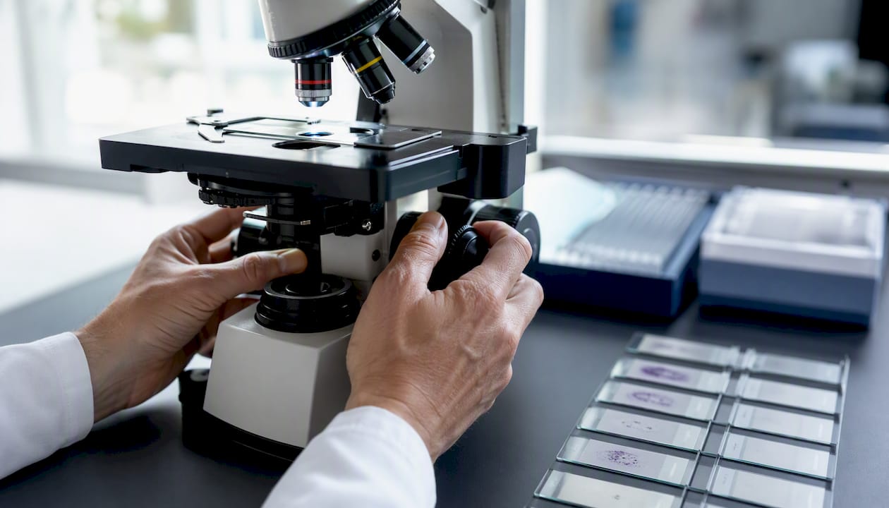
EIV Diagnostics, an independent pathology laboratory in Fresno, CA, offers dermatopathology services reviewed by board-certified dermatopathologists, alongside molecular pathology, digital pathology, and confocal microscopy. For providers and patients in the Central Valley, including Fresno, Clovis, Visalia, Hanford, and surrounding counties, the lab accepts both provider-ordered specimens and direct-to-consumer orders for patients who want to self-pay.
Pro Tip: When submitting a specimen, request that the requisition specify “dermatopathology review” and include the ABCDE features you or your provider observed. Labs that receive this context can prioritize the correct staining panel from the start, cutting days off the turnaround.
Why precision in diagnosis matters more than speed alone
The pressure to get results fast is understandable. But the more consequential variable is whether the report is complete the first time. A pathology report missing Breslow depth, ulceration status, or margin documentation is not a minor administrative gap. It can mean a second procedure, a delayed staging conversation, and weeks of uncertainty while the clinical team waits for an amended report.
The diagnostic steps outlined here, from dermoscopy through biopsy technique to molecular testing, exist because each one narrows uncertainty. Dermoscopy reduces unnecessary biopsies. Full-thickness excision gives the pathologist the tissue needed to measure depth accurately. A dermatopathologist review catches ambiguous lesions that a generalist might classify differently. Molecular testing on the same tissue block opens or closes treatment options without another procedure.
Patients who understand this sequence are better positioned to ask the right questions: Was this a full-thickness biopsy? Will a dermatopathologist review the slide? Does the report include ulceration status? Those questions are not demanding. They are exactly what guideline-aligned care looks like from the patient’s side.
Advanced diagnostic services through EIV Diagnostics
Patients and providers in the Central Valley who need dermatopathology, molecular pathology, or a second-opinion review have a direct path through EIV Diagnostics. The lab accepts specimens from provider orders, processes direct-to-consumer requests for patients with or without insurance, and offers mobile phlebotomy for specimen collection at home or in the office when a clinic visit is not practical.

For skin pathology specifically, EIV Diagnostics provides board-certified dermatopathology review, BRAF and other molecular panels run on biopsy tissue, and digital pathology consultation for complex or ambiguous cases. Providers can submit specimens with a standard requisition; patients can reach the lab directly to discuss self-pay options and skin pathology billing. If you need a second opinion on an existing pathology report, the lab can review slides and issue an independent interpretation. Contact EIV Diagnostics at Eivdiagnostics to start the process.
Sources
The following resources were used in preparing this article and are worth consulting directly for deeper reading:
- Diagnosis - Skin Cancer | FP Essentials
- Skin cancer: Symptoms, diagnosis, and causes
- Cutaneous Malignant Melanoma: Guideline-Based Management and Interprofessional Collaboration - StatPearls - NCBI Bookshelf
- Tests For Melanoma Skin Cancer | Melanoma Diagnosis | American Cancer Society
- Skin cancer - Diagnosis and treatment - Mayo Clinic
- Skin Cancer Screening (PDQ®)–Patient Version
This article is general information, not a substitute for advice from a qualified doctor. Consult a qualified healthcare professional about your own circumstances before acting on anything here.
FAQ
What does early-stage skin cancer look like on the face?
Early-stage skin cancer on the face often appears as a pearly or waxy bump (basal cell carcinoma), a flat flesh-colored or brown scar-like lesion, or a firm red nodule. Melanoma may present as a small pigmented spot with irregular borders or multiple colors. Any spot that changes, bleeds, or does not heal within a few weeks warrants evaluation.
Is skin cancer serious if caught early?
Most skin cancers caught at an early, localized stage are highly treatable. Basal cell carcinoma and squamous cell carcinoma have excellent outcomes with surgical removal. Early-stage melanoma (stage I) also carries a favorable prognosis, which is why prompt evaluation of suspicious lesions matters.
What is the survival rate for squamous cell carcinoma?
Squamous cell carcinoma of the skin has a high cure rate when caught early and treated with complete surgical excision. Localized SCC is associated with a 5-year survival rate above 90%; survival drops significantly when regional or distant spread is present, underscoring the importance of early diagnosis and complete margin removal.
How do you detect skin cancer early?
Monthly self-exams using the ABCDE criteria (asymmetry, border, color, diameter, evolving) help identify suspicious lesions early. Annual full-body skin exams by a dermatologist are recommended for people with risk factors such as fair skin, a history of sunburns, many moles, or a personal or family history of skin cancer. Dermoscopy during the clinical exam significantly improves the accuracy of early detection.
Recommended
- Dermatopathology Consultations: A Patient’s Guide | EIV Diagnostics | EIV Diagnostics
- Dermatopathology Services: What Patients and Providers Need to Know | EIV Diagnostics | EIV Diagnostics
- Skin Pathology Billing: A Clear Guide for Hanford Patients | EIV Diagnostics | EIV Diagnostics
- EIV Diagnostics | Independent Pathology Laboratory | EIV Diagnostics