
By EIV Diagnostics · July 8, 2026
Dermatopathology Consultations: A Patient's Guide
Discover how dermatopathology consultations can lead to accurate diagnoses for skin conditions. Learn what to expect and enhance your care.
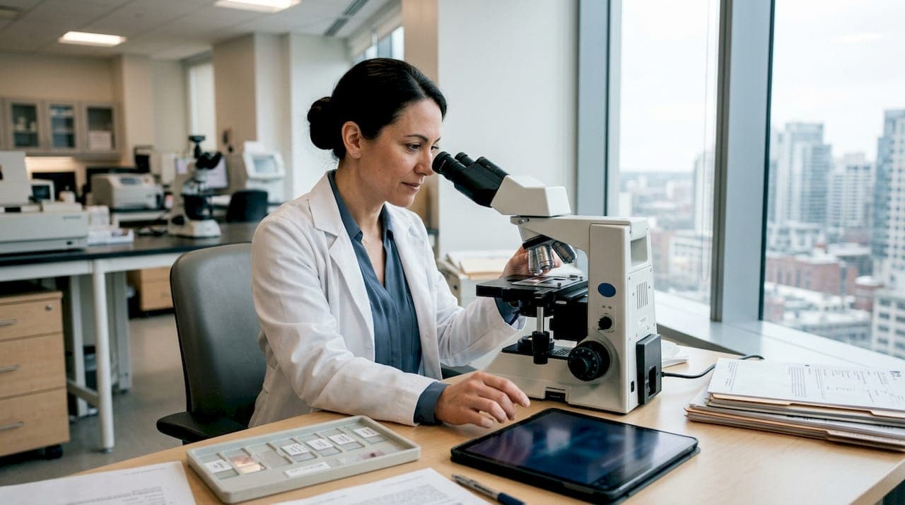
Dermatopathology consultations are specialized diagnostic services where expert physicians examine skin tissue samples under a microscope to deliver accurate diagnoses for a wide range of skin conditions. These consultations sit at the intersection of dermatology and pathology, requiring a level of training that takes 13 to 15 years to complete. For anyone in Fresno dealing with an unexplained rash, a suspicious mole, or a chronic skin condition that resists treatment, understanding how these consultations work puts you in a much stronger position. The right diagnosis is not just reassuring. It is the foundation of every effective treatment plan.
What are dermatopathology consultations and how do they work?
Dermatopathology consultations are defined as the expert review of skin biopsy specimens by a board-certified dermatopathologist. These specialists complete both dermatology or pathology residency training and a one-year fellowship, giving them a dual-path diagnostic perspective that neither a general pathologist nor a general dermatologist alone can replicate. Their job is to look at your skin tissue at the cellular level and determine exactly what is happening.
The term “dermatopathology” is the recognized clinical standard for this subspecialty. You may also hear it called cutaneous pathology or skin pathology consultation, but these all refer to the same process: a trained expert analyzing your biopsy to give your doctor a definitive answer.
What makes these consultations powerful is the combination of microscopic analysis and clinical context. A dermatopathologist does not just look at cells in isolation. They review your clinical history, the appearance of the lesion, and the biopsy findings together. That synthesis is what separates a confident diagnosis from a guess.
How do dermatopathologists diagnose skin conditions from biopsies?
The diagnostic process begins with a skin biopsy, and the type of biopsy your doctor chooses matters significantly. The three most common techniques are:
- Punch biopsy: A circular blade removes a small, full-thickness core of skin. This is the most common method for inflammatory conditions and suspected tumors.
- Shave biopsy: A blade skims a superficial layer of skin. Best suited for raised lesions like seborrheic keratoses or basal cell carcinomas.
- Excisional biopsy: The entire lesion is surgically removed. Used when melanoma or a deep lesion is suspected.
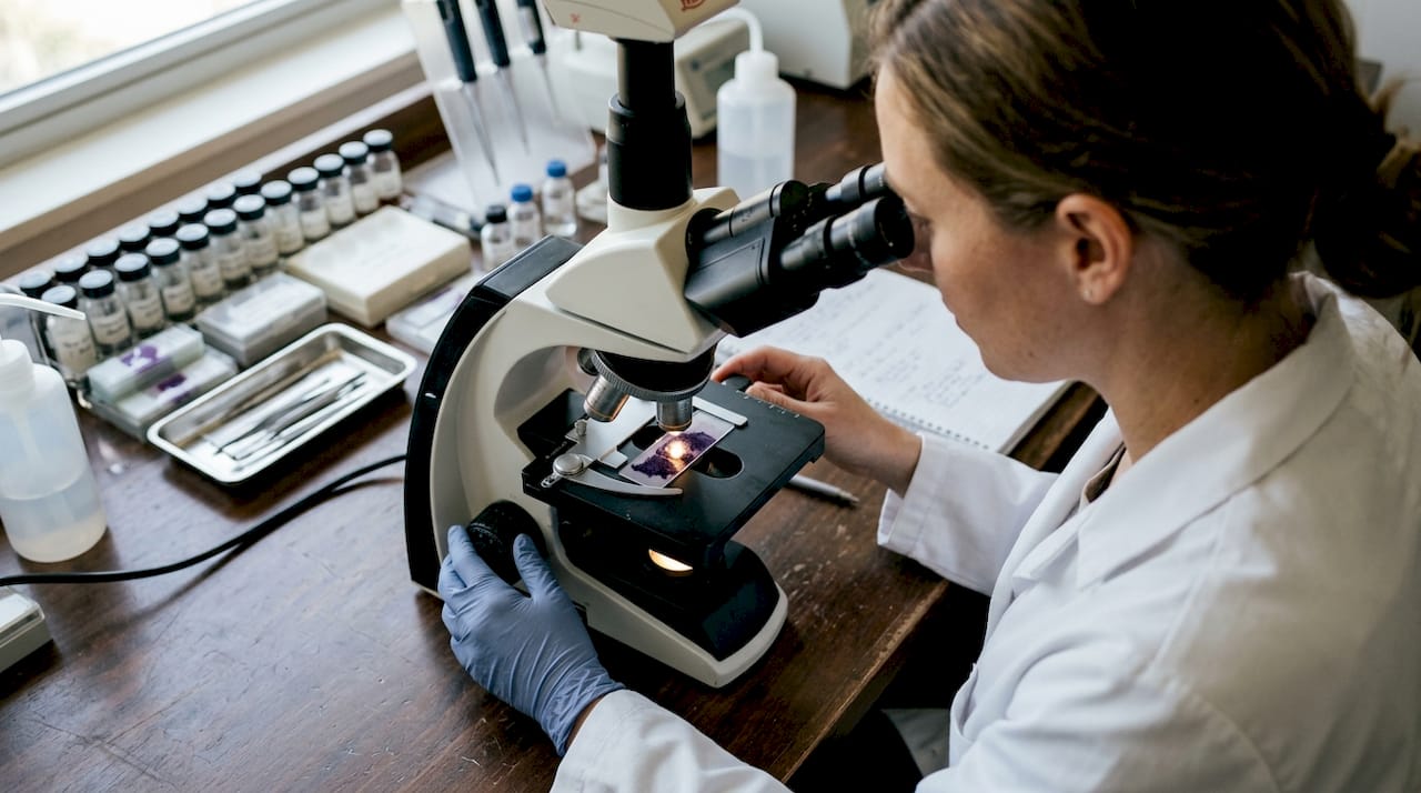
Once the tissue arrives at the laboratory, a dermatopathologist examines it under light microscopy. They assess cell shape, tissue architecture, inflammatory patterns, and any structural abnormalities. Routine light microscopy yields a specific histopathological diagnosis in approximately 80% to 83% of cases. That is a strong success rate, but it also means roughly 1 in 6 cases requires additional techniques.
Findings fall into broad categories: inflammatory conditions like psoriasis or eczema, neoplastic lesions ranging from benign cysts to melanoma, and rare conditions like vasculitis or cutaneous lymphoma. Most biopsy results are available within 2 to 4 weeks, though complex cases can take longer. Your clinical team will contact you once the analysis is complete and discuss next steps.
Pro Tip: For inflammatory rashes, ask your dermatologist about using a 4mm punch biopsy. Smaller samples can yield inconclusive results and lead to repeat procedures. Getting the right sample size the first time saves you time and discomfort.
When do dermatopathology consultations require specialized techniques?
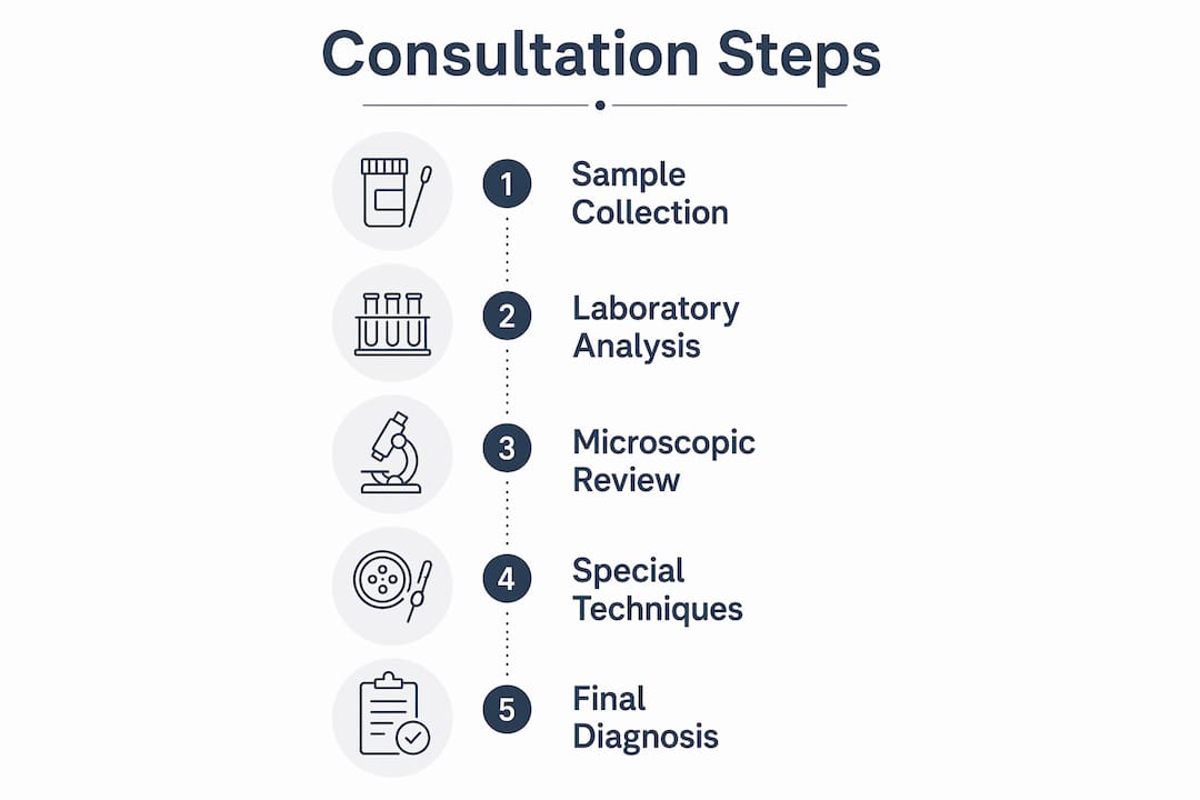
Routine light microscopy is powerful, but it has limits. When a biopsy does not produce a clear answer, dermatopathologists turn to specialized methods. Knowing which technique applies to which condition is a core part of their expertise.
| Technique | Primary indication | Diagnostic strength |
|---|---|---|
| Light microscopy | Most inflammatory and neoplastic conditions | Definitive in 80–83% of cases |
| Immunofluorescence | Autoimmune blistering diseases (pemphigus, bullous pemphigoid) | Identifies specific antibody deposits |
| Electron microscopy | Genetic disorders, storage diseases, ultrastructural defects | Reveals subcellular structures invisible to light microscopy |
| Molecular pathology (PCR) | Infectious agents, gene mutations, lymphoma subtyping | Provides definitive molecular identification |
Immunofluorescence is the go-to method for autoimmune blistering diseases. It uses fluorescent-labeled antibodies to detect where the immune system is attacking the skin. Without it, conditions like pemphigus vulgaris and bullous pemphigoid look nearly identical under standard microscopy.
Electron microscopy goes even deeper, revealing structures at the nanometer scale. It requires intensive sample preparation and specialized interpretation skills, which is why it is reserved for rare cases where light microscopy and immunostains are insufficient. Genetic skin disorders and certain storage diseases fall into this category.
Molecular pathology, including PCR-based testing, has expanded rapidly in clinical dermatopathology. It can identify specific infectious organisms, detect gene mutations linked to skin cancers, and subtype lymphomas with precision that no microscope alone can achieve. For patients with complex or rare diagnoses, these techniques are not optional extras. They are the path to a definitive answer.
What should you expect during and after a skin biopsy?
The biopsy procedure itself is straightforward. Most in-office biopsies take 15 to 20 minutes and involve local anesthesia to numb the area. Patients typically experience mild tenderness or bruising for 1 to 2 days afterward. Healing generally occurs within 7 to 14 days, depending on the biopsy site and technique used.
After the procedure, your tissue sample travels to a pathology laboratory for analysis. Here is what the post-biopsy period typically looks like:
- Days 1 to 3: Keep the biopsy site clean and covered. Mild soreness is normal.
- Days 4 to 14: The wound closes. Avoid submerging the area in water until fully healed.
- Weeks 2 to 4: Your pathology report is generated and reviewed by your clinical team.
- After results: Your dermatologist contacts you to discuss findings and any recommended next steps.
Patients should not interpret pathology reports independently. Diagnosis requires synthesizing microscopic findings with clinical examination data by a qualified physician. A report that mentions “atypical cells” or “mild dysplasia” means very different things depending on the full clinical picture.
Pro Tip: Before your follow-up appointment, write down three specific questions about your results. Ask what the diagnosis means for your daily life, what treatment options exist, and what signs would warrant a return visit. Prepared patients get more useful information from every appointment.
How can you use skin pathology consultations to navigate uncertain diagnoses?
Ambiguous diagnoses are more common than most patients realize. A lesion can show features of two different conditions, or a rare disease can mimic a common one. This is exactly where a dermatopathology second opinion delivers real value.
Direct consultations with dermatopathologists can help patients better understand complex diagnoses and reduce anxiety. These conversations go beyond the summary language in a written report. A dermatopathologist can explain what the cells looked like, why the diagnosis was reached, and what alternative possibilities were considered and ruled out.
Multidisciplinary collaboration also plays a major role in difficult cases. When a dermatopathologist, a treating dermatologist, and sometimes an oncologist review the same case together, the treatment plan that emerges is far more grounded. Each specialist brings a different lens, and the overlap is where diagnostic confidence is built.
Here are four steps you can take to engage proactively in your own diagnostic process:
- Request a copy of your pathology report. You have the right to this document. Reading it alongside your doctor helps you ask better questions.
- Ask whether a second opinion is appropriate. For rare conditions, ambiguous findings, or diagnoses that will lead to major treatment decisions, a second review is standard practice, not a sign of distrust.
- Provide complete clinical context. Tell your dermatologist about all medications, recent infections, family history, and the full timeline of your symptoms. This information directly affects how a dermatopathologist interprets your biopsy.
- Follow up if your symptoms change. A diagnosis is not always permanent. New symptoms can shift the clinical picture and warrant re-evaluation.
Taking these steps does not mean second-guessing your care team. It means participating in your own diagnosis, which consistently leads to better outcomes.
Key Takeaways
Dermatopathology consultations combine expert microscopic analysis with clinical context to deliver the definitive skin diagnoses that guide accurate, effective treatment.
| Point | Details |
|---|---|
| Expert training matters | Dermatopathologists complete 13–15 years of training, giving them unique diagnostic depth. |
| Biopsy type affects accuracy | Choosing the right biopsy technique and size, such as a 4mm punch for rashes, prevents inconclusive results. |
| Most cases resolve with light microscopy | Routine analysis yields a diagnosis in 80–83% of cases; specialized techniques handle the rest. |
| Second opinions are standard practice | For ambiguous or rare diagnoses, requesting a dermatopathology second opinion is appropriate and encouraged. |
| Patient preparation improves outcomes | Writing questions before appointments and providing full clinical history leads to more useful consultations. |
Why I believe dermatopathology is one of medicine’s most underappreciated specialties
Most patients never meet their dermatopathologist. The report arrives, the diagnosis is delivered, and the specialist who made the call remains invisible. That invisibility is a problem, not because dermatopathologists are doing anything wrong, but because patients who understand their diagnosis engage with treatment differently.
What I have observed working in diagnostic pathology is that the gap between a correct diagnosis and a useful diagnosis is communication. A report that says “superficial perivascular lymphocytic infiltrate consistent with early mycosis fungoides” is technically accurate. But a patient who understands that this means a rare type of lymphoma affecting the skin, that it is often slow-moving, and that it requires specific monitoring, is a patient who shows up to follow-up appointments and catches changes early.
The field is also evolving faster than most people realize. Molecular pathology techniques, including PCR and next-generation sequencing, are now part of routine dermatopathology workflows for complex cases. Digital pathology platforms allow specialists to review slides remotely with the same precision as in-person analysis. These advances mean that patients in Fresno have access to diagnostic depth that would have required a trip to a major academic center just a decade ago.
My honest advice: if you receive a skin diagnosis that will lead to surgery, long-term medication, or significant lifestyle changes, ask whether a dermatopathology consultation or second opinion has been completed. Not because your doctor is wrong. Because the stakes are high enough to be certain.
— Krunal
Eivdiagnostics offers expert skin pathology consultation in Fresno

Eivdiagnostics is an independent pathology laboratory based in Fresno, CA, with board-certified pathologists who specialize in dermatopathology, molecular pathology, and digital pathology. Whether your case involves a routine biopsy or a complex condition requiring PCR-based molecular analysis, Eivdiagnostics provides thorough analysis with fast turnaround times. The lab works directly with patients, providers, and self-pay customers, so access to expert diagnosis does not depend on navigating insurance barriers. Explore dermatopathology services at Eivdiagnostics, or review the full range of histopathology services available to patients and providers across the Central Valley.
FAQ
What is dermatopathology?
Dermatopathology is a medical subspecialty where trained physicians examine skin tissue samples under a microscope to diagnose skin diseases. It combines expertise from both dermatology and pathology to deliver cellular-level diagnoses.
How long does a skin biopsy result take?
Most skin biopsy results are available within 2 to 4 weeks. Complex cases involving specialized techniques like immunofluorescence or molecular testing may take longer.
When should I seek a dermatopathology second opinion?
A second opinion is appropriate when your diagnosis is rare, ambiguous, or will lead to major treatment decisions such as surgery or long-term medication. It is a standard part of responsible diagnostic care, not a challenge to your doctor’s judgment.
Can I meet directly with a dermatopathologist?
Direct consultations with dermatopathologists are possible and can help patients understand complex findings in plain language. These meetings reduce diagnostic anxiety and improve patient understanding beyond what a written report conveys.
Does biopsy size affect the accuracy of my diagnosis?
Yes. For inflammatory rashes, a 4mm punch biopsy provides enough tissue to distinguish between similar conditions. Smaller samples can produce inconclusive results and may require a repeat procedure.