
By EIV Diagnostics · September 26, 2026
Basal Cell Testing: 4 Biopsy Methods Confirming Diagnosis for Patients
Learn which biopsy confirms basal cell cancer, how labs analyze samples, and when special stains, imaging, or molecular tests actually change treatment.
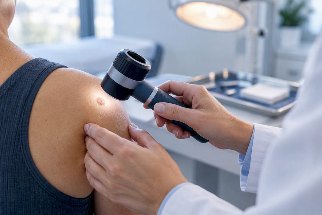
A biopsy examined by a pathologist is the test that confirms basal cell carcinoma. Clinical exam and dermoscopy come first and determine which biopsy technique to use, but neither can substitute for tissue under a microscope. Advanced molecular testing and imaging exist, but they play a supporting role reserved for specific, less common situations rather than standard first-line use.
TL;DR:
- Most diagnoses are confirmed through a skin biopsy, with shave and punch biopsies being the most common and least invasive methods.
- Dermoscopy significantly improves diagnostic accuracy before biopsy, especially by revealing patterns like arborizing vessels or pearly areas.
- Molecular testing is generally unnecessary for typical, localized BCC and is reserved for advanced or resistant cases due to its higher cost and longer turnaround times.
- Imaging is rarely needed unless there is suspicion of deep invasion, perineural spread, or recurrence in high-risk situations.
- Laboratory review by a dermatopathologist often provides clearer results, especially in ambiguous cases, and specialty labs like EIV Diagnostics enhance diagnostic precision and convenience.
Table of Contents
- What Is Basal Cell Testing and How Does the Process Start?
- Which Skin Biopsy Type Confirms Basal Cell Carcinoma?
- What Happens to the Tissue Sample in the Pathology Lab?
- Do You Need Molecular Testing or Special Stains for BCC?
- Is Imaging Ever Needed to Diagnose or Stage Basal Cell Carcinoma?
- How Long Do Basal Cell Testing Results Take and What Do They Mean?
- How Specialty Labs Improve Basal Cell Testing Accuracy and Convenience
- How Urgent Is Basal Cell Carcinoma Testing and Treatment?
- A Publisher’s View on Getting Basal Cell Testing Right
- Get Dermatopathology and Testing Support From EIV Diagnostics
- Sources
- FAQ
What Is Basal Cell Testing and How Does the Process Start?
Basal cell testing begins the moment a clinician looks closely at a suspicious spot, not in a lab. Before any biopsy tool touches the skin, a dermatologist or primary care provider works through a short set of questions and a visual exam designed to separate a probable basal cell carcinoma (BCC) from a mole, a scar, or a harmless bump.
Expect questions about how long the spot has been there, whether it has changed shape or color, whether it bleeds or crusts and comes back, and whether you have had skin cancer before. Sun exposure history and skin type matter too, since fair skin and years of unprotected sun exposure raise BCC risk substantially.
The visual exam usually involves a dermatoscope, a handheld magnifying device with polarized light that reveals patterns invisible to the naked eye. According to a 2024 review of BCC diagnosis, dermoscopy meaningfully improves diagnostic accuracy compared with a plain visual check, which is why most dermatologists use it routinely before deciding on a biopsy.
Specific patterns tend to show up under dermoscopy:
- Arborizing vessels: thin, branching blood vessels that look like tree limbs spreading across the lesion.
- Pearly or translucent areas: a glossy, almost waxy sheen that is a classic BCC signature.
- Ulceration: a central crater or open sore that will not fully heal.
- Blue-gray ovoid nests: rounded, bluish clusters seen more often in pigmented BCC subtypes.
None of these features alone proves cancer. A dermatologist evaluates the whole picture, and if suspicion stays high after the exam, standardized clinical photos often get taken to track the lesion or to send with a referral. At that point, the workup moves from looking to sampling.
Which Skin Biopsy Type Confirms Basal Cell Carcinoma?
Every path to a confirmed BCC diagnosis runs through a skin biopsy, but which technique gets used depends on the lesion’s size, location, and how confident the clinician already feels. There are four main approaches, and understanding the differences helps you know what to expect on the day of your procedure.
- Shave biopsy. A blade shaves off the top layers of skin down to about the depth of the dermis. This is the most common method for a suspected BCC because it is fast, requires only local anesthetic, and usually provides enough tissue for diagnosis. It leaves a shallow wound that typically heals with minimal scarring.
- Punch biopsy. A small circular tool, usually 3 to 4 millimeters wide, removes a cylindrical core of skin that goes deeper than a shave. Punch biopsies are preferred when the clinician needs to see the full thickness of the skin, which helps judge how deep a tumor might extend.
- Incisional biopsy. A wedge of tissue is cut from a larger lesion when removing the whole thing right away is not practical, often because of size or location near the eye, nose, or ear. This gives a diagnosis without committing to full removal on day one.
- Excisional biopsy. The entire visible lesion is removed with a margin of normal-looking skin around it. This is both diagnostic and, if margins are clear, potentially curative in one step. It requires more anesthetic, stitches, and a longer recovery than a shave or punch.
A partial biopsy, meaning a shave, punch, or incisional sample, is often adequate when the goal is simply to confirm what type of skin cancer is present before planning treatment. An excisional biopsy makes more sense when the lesion is small, in a low-risk location, and the clinician is confident enough to treat and diagnose at once.
Local anesthetic makes all four techniques far less uncomfortable than most patients expect. Aftercare is straightforward: keep the site clean and covered, expect some redness for a week or two, and follow whatever wound care instructions your clinician gives you. Scarring from a shave biopsy is usually faint. Excisional biopsies leave a linear scar that fades over months.
Pro Tip: If your lesion is on your face, ask your clinician directly why they chose a particular biopsy method. Dermatopathologists and dermatologists intentionally balance getting enough diagnostic tissue against leaving the smallest possible cosmetic mark, especially on visible areas.
There is also a same-day scenario worth knowing about. When Mohs micrographic surgery is planned, a biopsy confirming BCC can happen on the same day as the definitive surgical treatment, since Mohs relies on immediate histologic evaluation of tissue margins during the procedure itself. This requires a lab set up to read frozen sections in real time, which is a different workflow than standard biopsy processing.
What Happens to the Tissue Sample in the Pathology Lab?
Once a biopsy sample leaves the clinic, it enters a multi-step lab process before anyone can call it cancer. The tissue is first placed in a fixative solution, usually formalin, which preserves the cellular structure and stops decomposition. From there, it gets embedded in a paraffin wax block, sliced into sections just a few microns thick, and mounted on glass slides.
Those slides are stained, most commonly with hematoxylin and eosin (H&E), a combination that colors cell nuclei purple-blue and surrounding tissue pink. That contrast is what lets a pathologist actually distinguish tumor cells from normal skin under the microscope.
A general pathologist can identify obvious BCC, but subtle subtypes and tricky margins are where subspecialty training pays off. A dermatopathologist is a pathologist with focused training in skin disease, and that additional expertise matters most in ambiguous cases, mixed subtypes, or when a report needs to guide a decision as consequential as facial surgery. The AAD’s patient guidance notes that a dermatopathologist’s review provides the final diagnosis after the initial biopsy is taken.
A finished pathology report typically includes several standard elements:
- Diagnosis: confirmation of basal cell carcinoma or an alternative finding.
- Subtype: nodular, superficial, infiltrative, morpheaform, or micronodular, each with different growth behavior.
- Margin status: whether cancer cells reach the edge of the sampled tissue.
- Level of invasion: how deep the tumor extends into the dermis or beyond.
- Comments or recommendations: notes on whether further excision, additional stains, or a specialist referral would help.
Margin status deserves its own attention because it directly shapes what happens next. “Clear margins” means no tumor cells were found at the edges of the removed tissue, a reassuring sign, particularly after an excisional biopsy. “Positive margins” means cancer cells reach the border of the sample, signaling that more tissue may need to come out. An infiltrative or morpheaform subtype with positive margins often points toward Mohs surgery rather than standard excision, since these subtypes tend to spread in finger-like projections that are easy to underestimate on a single pass. Reading a pathology report can feel confusing on your own, which is exactly the kind of situation where a dermatopathology consultation or second opinion adds real clarity.
Do You Need Molecular Testing or Special Stains for BCC?
Most basal cell carcinomas never need anything beyond standard H&E staining. Ancillary tests get added only when the diagnosis is genuinely unclear or the tumor behaves in a way that demands more precision.

Immunohistochemistry (IHC) is the most common add-on. It uses antibodies tagged with visible markers to highlight specific proteins in the tissue, which helps a pathologist distinguish BCC from other skin cancers or benign growths that can look similar under a standard stain. For instance, BerEP4 staining helps separate BCC from squamous cell carcinoma when the architecture on a routine slide is ambiguous. These special stains resolve a genuine diagnostic question, not a routine formality.
Molecular and genomic testing sit at a different level entirely, and this is where expectations need to be realistic. FDA-cleared molecular and companion-diagnostic tools can analyze formalin-fixed tumor tissue for defined indications, but a broad genomic sequencing panel is not a routine first-line test for typical, localized BCC. A PMC review of current BCC diagnostic approaches notes that advanced molecular testing tends to matter most when a systemic therapy or clinical trial enrollment is on the table, not for garden-variety lesions caught early.
The clinical reality is straightforward: molecular testing earns its place when a tumor is advanced, recurrent, or resistant to standard treatment, not as a default add-on to every biopsy. Ordering it reflexively adds cost and turnaround time without changing the treatment plan for most patients.
Tissue conservation is part of the calculation too. Every slide cut for a special stain or molecular assay uses up sample that might otherwise be needed for further diagnostic review. Turnaround time stretches from a few added days for IHC to a couple of weeks for more complex molecular panels, and the price tag rises accordingly. If your clinician mentions molecular testing, a fair question to ask directly is whether the result would actually change your treatment plan. If the answer is no, you can reasonably skip it. Readers curious about how these tests get interpreted can look at a broader explanation of molecular testing for cancer and what results typically mean.
Is Imaging Ever Needed to Diagnose or Stage Basal Cell Carcinoma?
Imaging is the exception, not the rule, in basal cell carcinoma workups. BCC almost never spreads to distant organs, so the routine CT scans, MRIs, and PET scans used to stage other cancers simply are not part of a standard evaluation. The NCI’s PDQ summary on skin cancer treatment confirms that staging tests are typically unnecessary because BCC rarely metastasizes.
Imaging gets ordered only in specific, higher-risk situations:
- Suspected deep invasion: when a tumor appears to extend well beyond the skin into muscle, cartilage, or bone.
- Perineural spread: when cancer may be tracking along a nerve, which can cause numbness or shooting pain and needs mapping before surgery.
- Recurrence after prior treatment: especially when a tumor comes back in a scarred or previously irradiated area, making physical exam less reliable.
- Aggressive histology on the pathology report: infiltrative or morpheaform subtypes with concerning features sometimes prompt imaging to plan surgical extent.
The choice of scan follows the clinical question. MRI is the go-to for soft tissue detail and tracking perineural invasion, since it shows nerve involvement better than any other modality. CT scans are better suited to evaluating bone involvement. Ultrasound can check nearby lymph nodes when there is any concern about spread, though this is uncommon for BCC specifically. PET scans are rarely used, reserved for the rare advanced or metastatic case rather than typical disease.
How Long Do Basal Cell Testing Results Take and What Do They Mean?
Standard biopsy results for suspected basal cell carcinoma usually come back within a few days to about a week, once the sample completes fixation, processing, staining, and pathologist review. Add immunohistochemistry and that window stretches by several more days. Molecular testing, when it is genuinely indicated, can take one to two additional weeks depending on the specific assay and lab.
Turnaround times generally range from a few days for standard H&E biopsy review to additional days for immunohistochemistry and up to a couple of weeks for molecular or genomic panels when required.
Results typically get delivered to the ordering clinician first, who then calls or schedules a visit to walk through the findings with you. This is the conversation where report language actually gets translated into next steps, and it is worth asking questions rather than trying to decode medical terminology alone.
A few phrases show up often enough to deserve plain explanation. “Nodular BCC” describes the most common and generally slower-growing subtype. “Infiltrative” or “morpheaform” describes a subtype that grows in less predictable, finger-like patterns and often needs a wider surgical margin or Mohs surgery. “Positive margins” means the biopsy or excision did not fully capture the tumor’s edge, which usually leads to a second procedure. “Clear margins” is the reassuring version of the same finding. None of these terms mean the same thing for every patient, since location, subtype, and overall health all factor into what happens next.
How Specialty Labs Improve Basal Cell Testing Accuracy and Convenience
The quality of your basal cell testing outcome depends heavily on what happens after the biopsy leaves the exam room, and that is where a dedicated pathology lab makes a real difference. EIV Diagnostics is an independent pathology laboratory built around dermatopathology, molecular pathology, and digital pathology, with confocal microscopy available for cases that benefit from high-resolution cellular imaging.
Subspecialty review matters most in cases that are not textbook. A board-certified dermatopathologist is trained specifically to catch subtle subtype differences and margin nuances that a general pathologist might read differently. Digital pathology, which converts glass slides into high-resolution digital images, lets specialists consult on a case remotely without shipping physical slides across the country, which can shave real time off a diagnosis.
Here is what a patient or provider working with a specialty lab can reasonably expect:
- Clear, structured reports that spell out subtype, margin status, and depth of invasion rather than dense shorthand.
- Pathologist review on dermatopathology cases, with the option to request additional stains or a second opinion when something is ambiguous.
- Flexible ordering paths, since testing may be requested through a healthcare provider or, in some cases, directly by a patient through self-pay options.
- Mobile phlebotomy for blood-based companion testing, which may bring a phlebotomist to a home or office instead of requiring a separate lab visit.
Ordering logistics are simpler than most patients assume. A provider can send a biopsy sample directly with a written order, or a patient exploring options can look into self-pay testing arrangements for certain services. Turnaround expectations match the general ranges described earlier: days for standard histology, longer when additional stains or molecular work get added. The guiding principle stays the same whether you are working with any pathology lab: match the test to the actual clinical question. Ordering advanced testing without a clear reason wastes tissue, time, and money without improving your care.
How Urgent Is Basal Cell Carcinoma Testing and Treatment?
Basal cell carcinoma is rarely a medical emergency, but that does not mean it is safe to ignore. BCC grows slowly compared with melanoma, yet delay still allows a tumor to invade deeper tissue, which can mean more extensive surgery and a harder recovery later.
Certain signs should push you toward faster evaluation rather than a wait-and-see approach:
- Rapid growth over weeks rather than months.
- Persistent bleeding or a sore that will not heal after several weeks.
- Pain, tenderness, or numbness at the site.
- Location near the eye, nose, ear, or lip, where even modest growth can complicate treatment and reconstruction.
Most dermatology practices triage suspicious lesions within one to two weeks of a referral, faster if the presentation looks aggressive. If a lesion is caught early and looks classic for a low-risk subtype, waiting a couple of weeks for a scheduled appointment is not dangerous.
Pro Tip: While you are waiting for your appointment, protect the area from sun exposure and avoid picking or irritating it. Take a clear photo with a ruler or coin next to the lesion so you and your clinician have an objective size reference at the actual visit.
A Publisher’s View on Getting Basal Cell Testing Right
Too many patients treat a biopsy result as the end of the story instead of the beginning of a treatment conversation. The pathology report is where the real decisions get made, and a report that clearly names the subtype, margin status, and depth of invasion is worth far more than a fast turnaround with vague language. EIV Diagnostics built its dermatopathology and digital pathology services around that principle: board-certified review and reporting clear enough that a patient can actually understand what happened to their tissue, not just that “cancer was found.”
We think the biggest gap in basal cell testing is not access to advanced technology. It is the conversation between patient and clinician about what a given test will actually change. Molecular testing sounds impressive, but it does nothing for a straightforward nodular BCC caught early. A dermatopathologist’s careful read of a standard H&E slide does far more work in the vast majority of cases than any add-on test.
If your report leaves you with more questions than answers, ask for it explained line by line, and do not hesitate to request a specialist second opinion when the subtype or margins sound ambiguous. Testing exists to guide a decision, not to generate paperwork.
— EIV Diagnostics
Get Dermatopathology and Testing Support From EIV Diagnostics
Specialty pathology laboratories provide patients and providers a direct path to subspecialty dermatopathology review instead of routing suspicious skin biopsies through a general lab that may lack that focused expertise. That distinction shows up in report clarity: subtype, margin status, and depth of invasion spelled out in language that supports an actual treatment decision, not just a diagnosis code.

Services relevant to a basal cell workup include dermatopathology for biopsy interpretation, molecular pathology for the smaller subset of cases where genomic or companion testing is genuinely indicated, and digital pathology for faster specialist consultation on ambiguous slides. Providers can order testing directly, and patients without a standing order can explore self-pay testing options to move forward without waiting on insurance authorization.
For anyone who needs a blood draw as part of companion testing or a broader workup, EIV Diagnostics offers mobile phlebotomy starting at $65 per visit, bringing a phlebotomist to your home or office instead of requiring a separate trip to a draw site. If you have a biopsy result in hand and questions about what it means for treatment, reach out to EIV Diagnostics to ask about a dermatopathology consultation or a second review of your slides.
This article is general information, not a substitute for advice from a qualified doctor. Consult a qualified healthcare professional about your own circumstances before acting on anything here.
Sources
- Skin Cancer Treatment (PDQ®) - NCI
- Basal cell carcinoma: From symptoms to treatments - AAD
- One-Stop Shop: Diagnosis and Treatment of Basal Cell Carcinoma in One Step - PMC
- Billing and Coding: Mohs Micrographic Surgery (MMS) (A57767) - CMS
FAQ
What Are the Five Warning Signs of Basal Cell Carcinoma?
Common warning signs include a shiny or pearly bump, a sore that bleeds and does not heal, a flat scaly patch, a waxy scar-like area with no clear injury, and a pink growth with a slightly raised, rolled edge. Any lesion that changes shape, size, or color over weeks to months warrants an evaluation, since a dermatologist relies on these visual patterns alongside dermoscopy to decide on biopsy.
How Urgent Is It to Remove a Basal Cell Carcinoma?
BCC grows slowly and rarely spreads, so it is not usually a medical emergency, but delaying evaluation lets a tumor invade deeper tissue and complicate treatment. Most dermatology practices schedule suspicious lesions for biopsy within one to two weeks, sooner if the lesion is bleeding, growing rapidly, or located near the eyes, nose, or lips.
How Do Doctors Test for Basal Cell Carcinoma?
Doctors start with a visual exam and dermoscopy to assess suspicious features, then confirm the diagnosis with a skin biopsy read by a pathologist. A dermatopathologist’s microscopic review of the biopsy tissue provides the definitive diagnosis, and additional stains or molecular testing are added only in specific, less common cases.
What Does Stage 1 Basal Cell Cancer Look Like?
Early basal cell carcinoma often appears as a small, pearly or translucent bump, sometimes with visible fine blood vessels, or a flat pink to reddish patch that does not heal. Formal staging is uncommon for BCC because the disease rarely metastasizes, so most cases are managed based on tumor size, subtype, and location rather than a formal stage number.
Does EIV Diagnostics Offer Basal Cell Testing Services?
EIV Diagnostics provides dermatopathology review for skin biopsies, including basal cell carcinoma cases, along with molecular pathology and digital pathology for cases that need additional analysis. Pricing for most diagnostic services is not published; readers can check current details directly on the EIV Diagnostics site or inquire about self-pay options.