
By EIV Diagnostics · November 9, 2026
Lichen Planus Pathology: What the Biopsy Shows Under the Microscope
Lichen planus histology explained: the classic biopsy findings, how oral and variant forms differ, look-alikes, and what to expect when your skin biopsy is read.
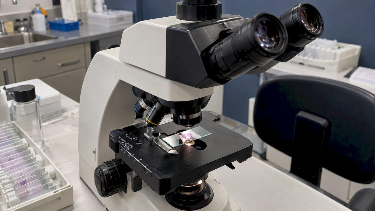
Lichen planus pathology comes down to one recognizable pattern: a lichenoid interface dermatitis. Under the microscope, a pathologist sees a thickened outer skin layer, a dense band of lymphocytes hugging the junction between epidermis and dermis, damaged cells at the base of the epidermis, and a jagged, sawtooth profile to the rete ridges. Pathology Outlines describes lichen planus clinically by the “6 Ps” (purple, pruritic, planar, polygonal papules and plaques) and histologically by that interface pattern. This guide walks through each feature, how the oral and variant forms differ, what mimics lichen planus, and how a good biopsy is taken and read.
TL;DR
- The classic picture is hyperkeratosis, wedge-shaped hypergranulosis, sawtooth rete ridges, basal cell damage, and a band-like lymphocytic infiltrate in the upper dermis.
- Civatte (cytoid) bodies are dead keratinocytes. They are a helpful clue but are not seen in every case.
- Oral, hypertrophic, pigmentosus and atrophic forms each shift the picture slightly, so the clinical site and appearance matter.
- Lichenoid drug reactions, lupus, and a few other conditions can look similar. Eosinophils, follicular involvement and clinical history help separate them.
- A fresh, untreated lesion and the right biopsy site make the report more useful.
Table of Contents
- What is lichen planus, and what does the pathologist examine?
- The classic histologic features of lichen planus
- How histology differs by type of lichen planus
- What else can look like lichen planus?
- Where direct immunofluorescence fits
- How the biopsy is taken matters
- Reading your pathology report
- Our perspective
- Dermatopathology support in Fresno and the Central Valley
- FAQ
- Sources
What is lichen planus, and what does the pathologist examine?
Lichen planus (LP) is a chronic, immune-mediated inflammatory condition that affects skin, mucous membranes, nails and hair. A 2025 review reports it affects about 0.5% to 1% of the population in the literature it surveys. On the skin it typically appears as itchy, flat-topped, purplish, polygonal bumps. Many clinicians can suspect it at the bedside, but the diagnosis is usually confirmed with a biopsy.
The clinical guide published in JDDG recommends that the clinical diagnosis be confirmed by histopathology, because the classic band-like pattern is often enough to rule out other disorders. The pathologist receives a small piece of skin, processes it into thin stained slides, and reads the architecture from the surface down to the upper dermis.
The classic histologic features of lichen planus
A well-developed, active lesion of cutaneous LP typically shows the following, according to Pathology Outlines and an academic update on LP and its variants.
Surface and epidermis
- Hyperkeratosis. The stratum corneum is thickened, usually without nuclei retained (orthokeratosis).
- Wedge-shaped hypergranulosis. The granular layer is thickened in a focal, wedge-like pattern. This is the microscopic counterpart of Wickham’s striae, the fine white lines seen on the surface of a papule, as noted in a 2023 clinicopathologic study.
- Sawtooth acanthosis. The rete ridges become pointed and irregular, like the teeth of a saw. This profile is one of the most recognizable low-power clues.
- Basal layer damage. Vacuolar change and apoptotic keratinocytes appear at the base of the epidermis.
The junction and dermis
- Band-like lymphocytic infiltrate. A dense, sharply bordered band of lymphocytes fills the upper dermis and can obscure the dermoepidermal junction. Most of these cells are memory T cells, identified with CD8 and CD45RO markers.
- Civatte (cytoid) bodies. These are necrotic keratinocytes, roughly 20 μm across, with homogeneous pink (eosinophilic) cytoplasm. In a 38-case analysis, they can lie in clusters in the uppermost dermis.
- Pigment incontinence. Melanin may drop into the dermis and be taken up by macrophages, especially in older lesions.
How consistent are these features?
Not every feature shows up in every slide. In the 38-case series, hyperkeratosis was present in 92%, hypergranulosis in 82%, and basal cell vacuolization in 100%, while the band-like infiltrate appeared in every case and Civatte bodies in 29%. A 155-case study from central India found hyperkeratosis in 99.35% of cases, wedge-shaped hypergranulosis in 65.80%, and a band-like dermal infiltrate in 74.87%. The takeaway: the pathologist weighs the whole pattern, not any single finding.

How histology differs by type of lichen planus
The core lichenoid reaction stays the same across forms, and an academic review notes that light microscopy shows similar features even when a rare variant looks very different on the skin. Still, there are useful differences, summarized from Pathology Outlines.
| Form | Typical clinical look | Histologic emphasis |
|---|---|---|
| Classic cutaneous | Purple, flat-topped, itchy papules | Hyperkeratosis, wedge-shaped hypergranulosis, sawtooth rete ridges, band-like infiltrate |
| Oral | White lacy or erosive patches | Occasional parakeratosis, less acanthosis than skin, plasma cells in the infiltrate |
| Hypertrophic (hyperkeratotic) | Thick, scaly plaques | Striking hyperkeratosis and acanthosis, pseudoepitheliomatous hyperplasia, sometimes eosinophils |
| Pigmentosus | Dark brown macules and papules, often without symptoms | Moderate lymphocytic infiltrate with dermal melanin incontinence |
| Atrophic | Flat, thinned lesions | No acanthosis, fewer Civatte bodies, more pigment incontinence, minimal inflammation |
Hyperkeratotic (hypertrophic) lichen planus
This form produces thick plaques, often on the shins. Under the microscope the epidermis is markedly thickened, and the overgrowth can mimic a squamous proliferation, which is why the term pseudoepitheliomatous hyperplasia appears in reports. A 2023 pediatric case report of linear hypertrophic LP documented hyperkeratosis, papillomatosis, wedge-shaped hypergranulosis, and a band-like infiltrate with basal vacuolar change. In the larger Indian series, hypertrophic LP made up 29.6% of cases, close behind the classic form at 30.96%.
Lichen planus pigmentosus
Lichen planus pigmentosus presents with dark brown macules and papules that are often asymptomatic. The infiltrate is milder, and the standout feature is melanin in the dermis. The clinical story (location, skin type, duration) helps the pathologist interpret a quieter slide.
Oral and mucosal lichen planus
On mucosal surfaces, the epithelium is thinner and the sawtooth and wedge features can fade. Per a vulvar dermatoses review, mucosal LP may show parakeratosis and a thinned epithelium. Interpretation depends on good communication with the clinician. A Modern Pathology review points out that oral lichenoid drug reactions look nonspecific, so the report relies on medication history and what happens when a drug is stopped.
What else can look like lichen planus?
Several conditions produce a lichenoid pattern. Pathology Outlines lists lichenoid drug reaction, benign lichenoid keratosis, chronic discoid lupus, secondary syphilis and a keratoacanthoma-type squamous cell carcinoma among the look-alikes.
- Lichenoid drug reaction. Eosinophils in the infiltrate favor a drug reaction over idiopathic LP, though they can occur in hypertrophic LP. A 2024 retrospective study also lists parakeratosis, focal hypogranulosis, acanthosis and pigment incontinence as helpful clues.
- Discoid lupus erythematosus. Atrophy of the epidermis, follicular plugging and involvement of hair follicles and adnexa point toward lupus. Direct immunofluorescence shows granular deposits at the basement membrane in about 70% of discoid lupus samples, per the Modern Pathology review.
- Keratoacanthoma and squamous cell carcinoma. Thick hypertrophic LP can be confused with these. Our article on keratoacanthoma pathology covers how that lesion is separated from squamous cell carcinoma.
Because the overlap is real, the clinical information on the requisition form matters: where the lesion is, how long it has been present, and the patient’s medication list.
Where direct immunofluorescence fits
Most LP cases are settled on routine stains. Direct immunofluorescence (DIF) is a second tool for unclear cases. The 38-case analysis says DIF may help when features are non-specific or overlap with diseases such as discoid lupus. In LP it often shows numerous IgM-staining cytoid bodies in the dermal papillae or near the basal layer, as described in the update on lichen planus.
The pattern is not diagnostic on its own, because fibrinogen, IgM and complement can also appear in lupus and erythema multiforme. It supports the diagnosis only alongside the H&E findings and the clinical picture. For clinicians, our guide to skin biopsy types and DIF tips explains how to avoid repeat biopsies.

How the biopsy is taken matters
The quality of the sample shapes the quality of the answer. Guidance from the JDDG clinical guide says to take the biopsy from a fresh and, if possible, untreated lesion. For oral LP, take it as far as possible from mechanical stress and avoid the chewing area.
Two more points apply to mucosal disease:
- The oral diagnosis requires evaluating the basement membrane zone, so the biopsy needs intact, full-thickness epithelium. A sample of only the ulcerated part will not show the interface changes.
- For erosive LP, sample the edge of an eroded area, as the vulvar dermatoses review advises.
If you are a patient, your clinician chooses the site and technique. You can help by telling them about every medication and supplement you take and by mentioning any treatments you have already used on the area. Our patient guide to dermatopathology consultations explains what happens after the sample leaves the office.
Reading your pathology report
Reports vary in wording, but you will often see phrases like “lichenoid interface dermatitis,” “consistent with lichen planus,” or “lichenoid dermatitis, differential includes lichenoid drug reaction.” The last version is not a failure to diagnose. It means the microscope shows the pattern, and the final label depends on your history. Bring the report to the clinician who ordered the biopsy, since they know your symptoms and medications. Questions about treatment belong with your dermatologist, and oral or genital symptoms deserve a follow-up with the right specialist for that site.
Our perspective
We read lichen planus slides every day as part of our dermatopathology work, and our experience is that the diagnosis rests on three things together: a well-chosen biopsy, a careful read of the whole pattern, and the clinical context.
At EIV Diagnostics, board-certified pathologists with subspecialty expertise review skin specimens, and we aim for rapid turnaround so clinicians can act on the result. As a CAP-accredited laboratory, we hold ourselves to defined quality standards in how specimens are processed and reported.
We encourage referring clinicians to send a short history, a medication list and the lesion site with every specimen. That small step lets our pathologists weigh the look-alikes, such as lichenoid drug reaction or lupus, and write a report that answers the question you actually asked.
EIV Diagnostics
Dermatopathology support in Fresno and the Central Valley
EIV Diagnostics is an independent pathology laboratory in Fresno, CA, serving patients and healthcare providers across Fresno, Clovis, Madera, Visalia and the surrounding Central Valley. Our dermatopathology services cover skin biopsy interpretation, backed by histopathology and digital pathology tools.

If you want lab work without a visit to a clinic, our mobile phlebotomy brings the blood draw to your home or office, from $65. We also accept self-pay orders through our self-pay testing page. Providers can start with our services overview.
FAQ
What is the most characteristic histologic feature of lichen planus?
There is no single one, but the combination of a band-like lymphocytic infiltrate at the dermoepidermal junction, sawtooth rete ridges and wedge-shaped hypergranulosis is the hallmark. Basal cell damage with Civatte bodies adds support. Pathologists weigh the whole pattern.
What is hyperkeratotic lichen planus?
It is also called hypertrophic lichen planus, and it forms thick, scaly plaques. Histologically it shows striking hyperkeratosis and acanthosis with pseudoepitheliomatous hyperplasia, and sometimes eosinophils. Its look can overlap with other thick lesions, so biopsy confirmation is important.
What is lichen planus pigmentosus?
It causes dark brown macules and papules that are often without symptoms. Under the microscope there is a moderate lymphocytic infiltrate and melanin in the dermis (melanin incontinence), as Pathology Outlines describes.
Can a biopsy tell lichen planus from a drug reaction?
Sometimes it points the way. Eosinophils, parakeratosis and focal hypogranulosis favor a lichenoid drug reaction. Because findings can overlap, the pathologist also needs your medication timeline, and your clinician may track whether the rash settles after a drug is stopped.
Is a biopsy always needed for lichen planus?
Not always, since many cases are recognized clinically. But a clinical guide recommends histopathologic confirmation, and it matters most for atypical, thick, or mucosal lesions. Your dermatologist decides when a biopsy is warranted.
Sources
- Pathology Outlines: Lichen planus
- Update on lichen planus and its clinical variants
- Lichen Planus: A Clinical and Immuno-Histological Analysis
- Clinical and histopathological study of cutaneous lichen planus
- DermNet NZ: Dermatopathology, inflammatory skin diseases
- Lichen planus: a clinical guide (JDDG)
- Oral lichenoid lesions: distinguishing the benign from the malignant
- Vulvar Dermatoses: A Review and Update
- Lichenoid drug reactions: a retrospective study
- A Case of Linear Hypertrophic Lichen Planus in a Pediatric Patient
- Overview of lichen planus pathogenesis and review