
By EIV Diagnostics · August 22, 2026
What Confocal Microscopy Does for Skin Diagnosis
Discover how confocal microscopy enhances skin diagnosis by providing real-time, noninvasive imaging for accurate cancer detection and treatment.
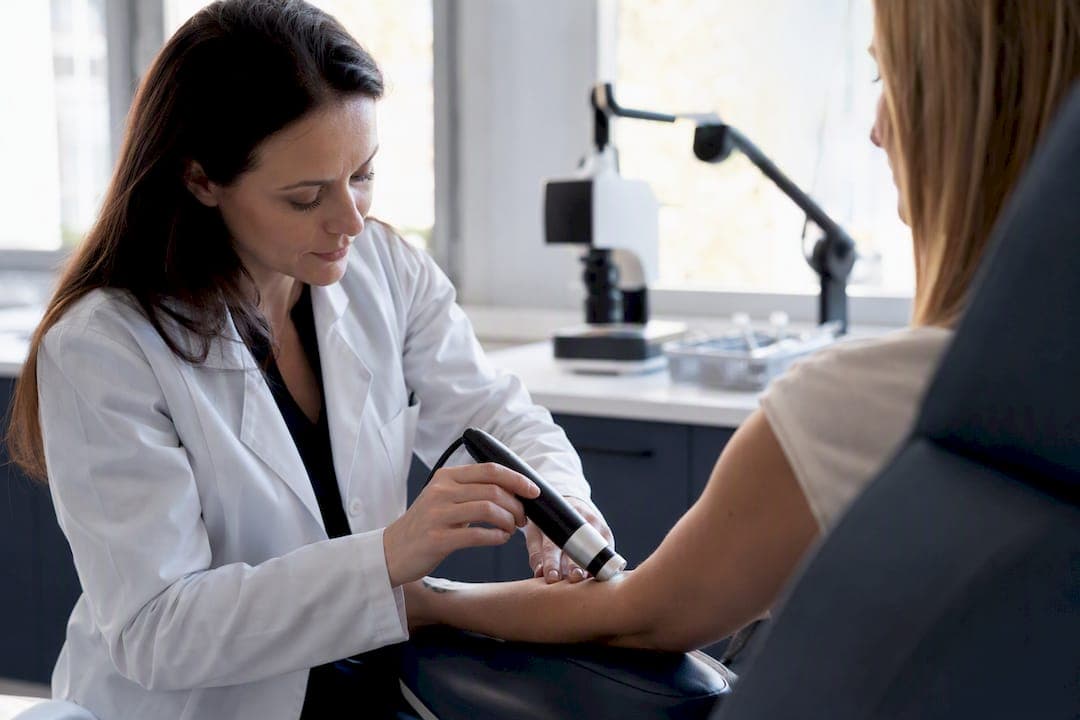
Reflectance confocal microscopy (RCM) is a noninvasive imaging test that lets clinicians view skin at near-histologic resolution, in real time, without cutting into it. It works as an optical biopsy: light from a low-power laser bounces off skin structures and builds a cellular-level picture of the epidermis and upper dermis, layer by layer.
Meta-analyses put pooled sensitivity for detecting skin cancers around 93.6% and specificity around 82.7%, and RCM imaging combined with dermoscopy has been shown to cut down on unnecessary excisions compared to dermoscopy alone. In the United States, CMS assigned CPT reimbursement codes for RCM acquisition and interpretation back in 2016, which means this isn’t an experimental curiosity sitting in a handful of academic centers. It’s billable, and increasingly standard, at least at the sites that own the equipment.
The catch is depth. RCM images the epidermis and superficial dermis reliably but loses resolution past roughly 200 to 300 micrometers, so it cannot substitute for histopathology when deeper invasion is in question.
- What it is: a laser-based, noninvasive imaging technique producing near-histologic, en face images of skin
- What it’s good for: triaging equivocal pigmented and nonpigmented lesions, guiding biopsy site selection, monitoring treatment response
- What it can’t do: assess deep dermal or subcutaneous disease, replace tissue diagnosis when malignancy is confirmed
Quick stat: Pooled diagnostic accuracy across skin cancer types runs close to 93.6% sensitivity and 82.7% specificity, with melanoma-specific sensitivity near 92.7%, according to a widely cited meta-analysis on RCM’s clinical role.
Key Takeaways
RCM works because it gives clinicians near-histologic, cellular-level images of skin without a scalpel, and its real strength is targeting biopsies precisely rather than replacing tissue diagnosis outright.
| Point | Details |
|---|---|
| Depth is the hard ceiling | RCM images reliably to about 200 to 300 micrometers, so deep dermal or subcutaneous disease needs a different test. |
| Performance is strong but not absolute | Pooled sensitivity near 93.6% and specificity near 82.7% support RCM as a triage tool, not a standalone diagnosis. |
| Add it after dermoscopy, not instead of it | Reserve RCM for equivocal lesions where dermoscopy alone leaves real ambiguity. |
| Billing is already established | CMS granted CPT codes for acquisition and interpretation in 2016, making RCM a billable clinical service. |
| Interpretation skill is the real bottleneck | Pattern-recognition training matters more than probe type for accurate reads. |
| EIV Diagnostics supports the tissue side | Board-certified pathologists at EIV Diagnostics provide dermatopathology correlation when RCM findings need histologic confirmation. |
Table of Contents
- How Confocal Microscopy Skin Imaging Actually Works
- Where Confocal Microscopy Dermatology Applications Deliver Real Value
- What Happens During an RCM Exam
- When Confocal Microscopy Skin Imaging Falls Short
- Building RCM Into a Dermoscopy-First Workflow
- Reimbursement and Coding for RCM in the United States
- Confocal Imaging Services Through EIV Diagnostics
- Training and Certification for Clinicians Using RCM
- What Confocal Microscopy Actually Costs a Practice
- Confocal Microscopy Versus OCT and Ultrasound for Skin
- Where Confocal Skin Imaging Is Headed Next
- Editorial Take: What Actually Matters With RCM
- Get Confocal Imaging and Dermatopathology Support From EIV Diagnostics
- Sources
- FAQ
How Confocal Microscopy Skin Imaging Actually Works
RCM relies on the same basic principle as any confocal system: a pinhole aperture rejects out-of-focus light so only a thin optical slice reaches the detector. In skin imaging, the light source is typically a near-infrared diode laser around 830 nanometers, chosen because it penetrates tissue reasonably well while staying under safety thresholds for continuous skin exposure. Contrast comes from differences in refractive index between cellular structures, mainly melanin, keratin, and collagen, rather than from injected dye or stain.
Resolution is the reason dermatologists call this a “virtual biopsy.” Lateral resolution runs about 0.5 to 1 micrometer and axial resolution about 3 to 5 micrometers, fine enough to distinguish individual keratinocytes, melanocytes, and even nuclear detail in some cases, according to the same PMC clinical review. Imaging depth tops out around 200 to 300 micrometers, which typically covers the epidermis and papillary dermis, per DermNet NZ’s technical overview.
Two probe designs dominate clinical practice, and the choice matters for workflow:
- Wide-probe (mosaic) systems, such as the class of devices built around a fixed stage, capture large mosaic fields by stitching together hundreds of individual frames. They’re excellent for flat, accessible sites like the trunk or extremities but awkward on curved or mobile surfaces.
- Handheld probes trade some field size for maneuverability, making them the better option for the face, ears, and other anatomically tricky areas where a rigid stage won’t sit flush.
Image output comes in a few flavors clinicians need to recognize. A mosaic is a wide-field composite at a single depth, useful for spotting architectural disorder across a lesion. A stack is a series of images taken at increasing depths from the same spot, letting you trace a structure like a nest of melanocytes as it descends toward the dermal-epidermal junction. Some newer platforms bundle stacks into volumetric “cubes” for 3D review. All of these are oriented en face, meaning parallel to the skin surface, which is a different plane than the vertical sections pathologists read on an H&E slide, and it takes deliberate practice to translate one into the other mentally.
A related but distinct technique, ex vivo fluorescence confocal microscopy, images freshly excised tissue rather than intact skin. It’s used mainly for rapid intraoperative margin checks during Mohs surgery, while in vivo RCM remains the workhorse for noninvasive diagnosis and monitoring.
Where Confocal Microscopy Dermatology Applications Deliver Real Value
The clinical case for RCM rests on numbers that hold up across multiple pooled analyses, not on isolated case reports. For melanoma specifically, sensitivity runs about 92.7% and specificity closer to 78.3%, according to pooled data from clinical studies. Those numbers matter less as abstract percentages and more for what they imply: RCM is strong at ruling lesions in or out of a suspicious category, but it’s a triage layer, not a replacement for tissue diagnosis when the picture stays ambiguous.
For melanoma, readers look for pagetoid spread (single atypical melanocytes scattered through the epidermis, appearing bright against surrounding keratinocytes), disarray of the normal honeycomb keratinocyte pattern, and nonedged dermal papillae. Cerebriform nests at the dermal-epidermal junction are another classic tell.
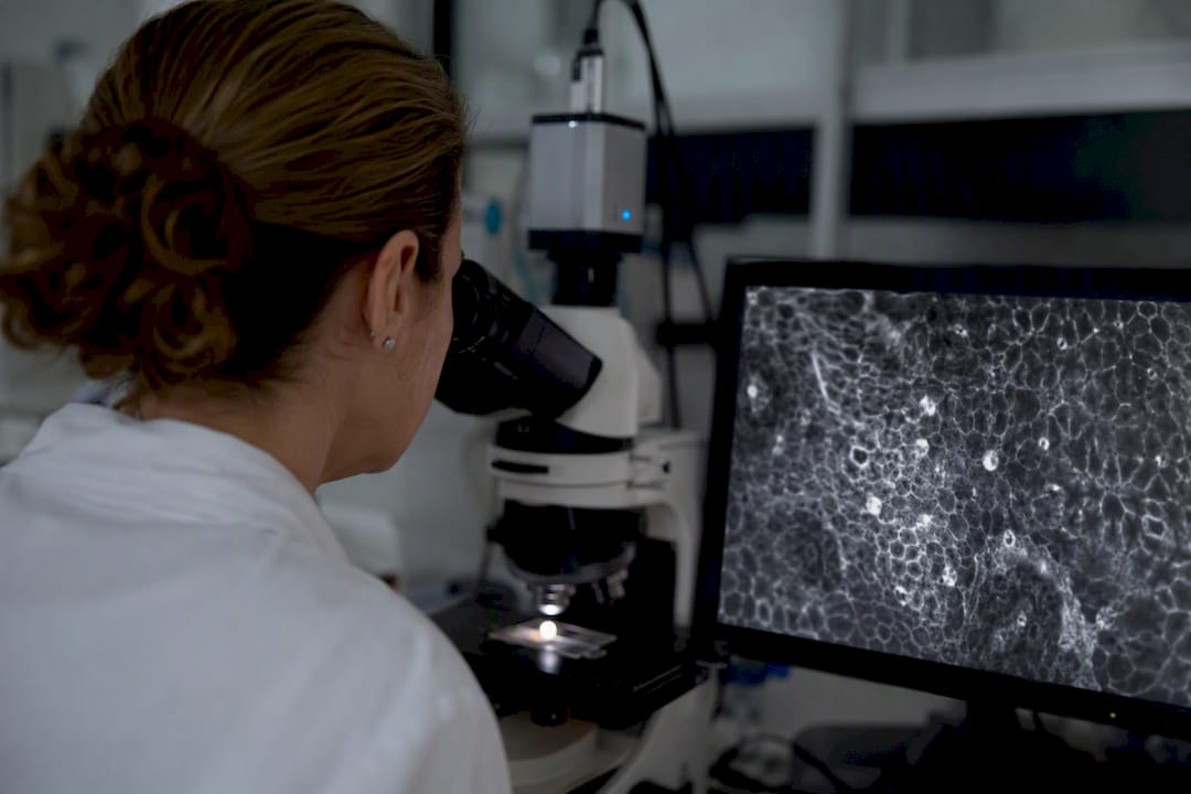
For basal cell carcinoma, the signature findings are elongated monomorphic nuclei polarized along the same axis (often described as “streaming”), dark silhouettes corresponding to tumor islands, and increased dermal vascularity feeding the tumor.
For squamous cell carcinoma and actinic keratosis, expect architectural disruption of the granular and spinous layers, atypical honeycombing, and round nucleated cells at the spinous layer replacing the normal pattern.
Beyond oncology, RCM has a growing footprint in inflammatory dermatology. It can visualize spongiosis in eczema, characterize fungal hyphae in tinea infections without waiting on a culture, and track how a psoriatic plaque responds to topical therapy over weeks, giving objective evidence for a treatment change instead of a subjective clinical impression alone.
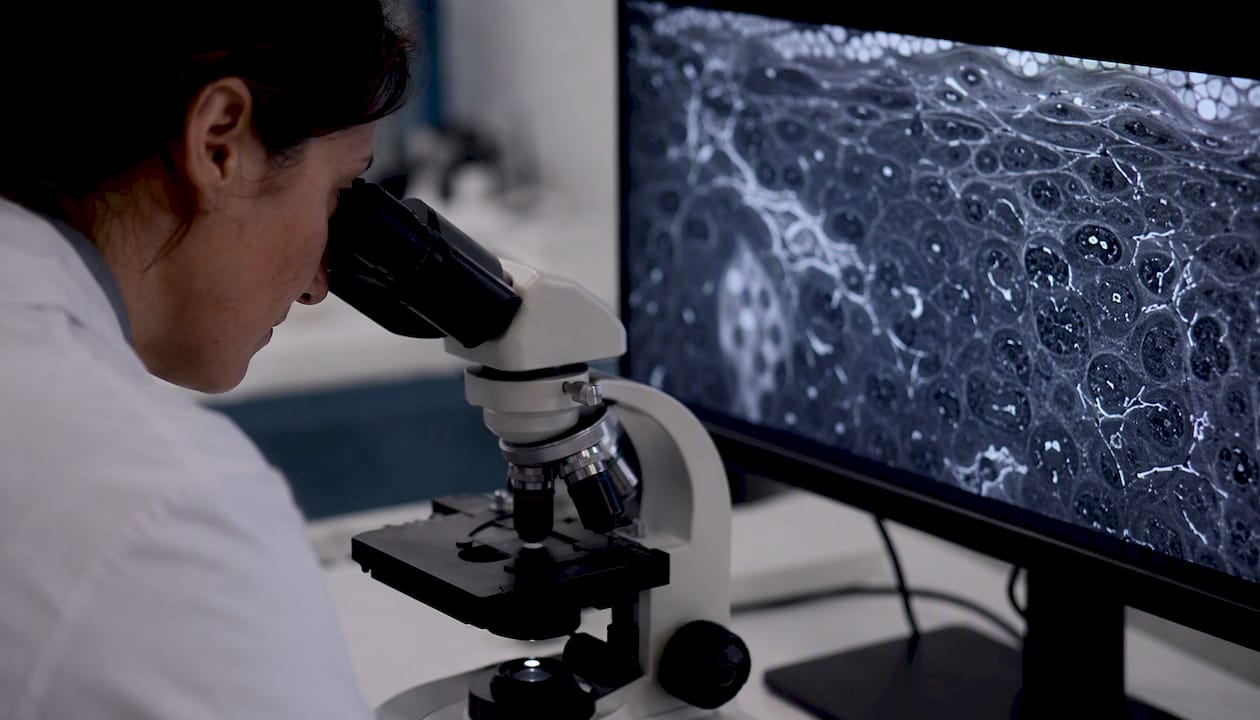
One of the more underappreciated uses is presurgical margin mapping. Before Mohs surgery or a wide local excision, RCM can scan the periphery of a lesion, particularly a lentigo maligna with an indistinct clinical border, and flag subclinical extension that would otherwise be missed until final pathology. That single application has changed how some surgeons plan their first cut, especially on cosmetically sensitive sites like the central face where every millimeter of margin counts. Adding RCM to dermoscopic evaluation has been shown to increase specificity meaningfully on sun-damaged skin, where dermoscopy alone tends to overcall biopsies because of background solar changes.
What Happens During an RCM Exam
Patients don’t need to do much to prepare, which is part of the appeal. Skip lotions, sunscreen, or heavy makeup on the area being imaged that day, since residue can scatter the laser and blur the mosaic. If the lesion sits under thick terminal hair, the clinician may trim it beforehand; visible scale or crust sometimes gets gently debrided first, since keratin blocks the laser almost as effectively as it blocks visible light.
- Site marking and cleaning. The lesion is identified, sometimes photographed with dermoscopy first for correlation, and the skin surface is wiped clean.
- Window cap placement. A small adhesive or metal ring, sometimes called a tissue window, is applied to the skin to stabilize the area and prevent motion artifact during scanning.
- Immersion medium application. A drop of gel or oil fills the space between the probe and the skin, matching refractive indices so light transmits cleanly instead of scattering at the surface.
- Mosaic and stack acquisition. The operator captures a wide-field mosaic at the dermal-epidermal junction, then takes stacks at multiple depths through areas of interest.
- Immediate review. Images display in real time, so the clinician can decide on the spot whether to biopsy, monitor, or reassure the patient, a workflow described in Memorial Sloan Kettering’s patient-facing guidance.
Total appointment time runs 15 to 45 minutes depending on lesion size and how many sites need imaging, per DermNet NZ. The laser is nonionizing and there’s no needle, so most patients describe the sensation as mild pressure from the probe, nothing more. Facial and trunk lesions image well; palms, soles, and the nail unit are harder because thick stratum corneum or nail plate blocks laser penetration.
Pro Tip: Book RCM early in a lesion’s course rather than after it has thickened or crusted over. Hyperkeratotic and ulcerated lesions scatter the laser before it reaches the dermal-epidermal junction, which is exactly the layer you need to see.
When Confocal Microscopy Skin Imaging Falls Short
RCM’s biggest structural limitation is depth. Once a lesion invades beyond about 200 to 300 micrometers, you’re imaging blind, and there’s no way to confirm or exclude deeper dermal involvement without tissue. That makes RCM a poor standalone tool for anything you already suspect is invasive on clinical grounds.
Several practical factors degrade image quality even within that depth range:
- Hyperkeratosis and scale scatter and absorb the laser before it reaches deeper layers, common in actinic keratoses and some squamous lesions.
- Motion artifact from breathing, pulse, or patient discomfort blurs stacks, particularly on the trunk near the ribs.
- Probe pressure applied unevenly can blanch the skin, compressing vasculature and distorting the very features you’re trying to assess.
- Concave or highly curved sites, like the nasal ala or ear helix, make it hard to maintain even contact with a wide-probe system, often forcing a switch to handheld.
The interpretation gap deserves equal attention. Reading RCM mosaics and stacks accurately requires deliberate training in pattern recognition, distinguishing pagetoid cells from artifact, tumor islands from normal adnexal structures, edged versus nonedged papillae, according to clinical guidance on RCM interpretation. A novice reader can produce false reassurance just as easily as a false alarm, and there’s no substitute for volume and mentored feedback while building that skill.
On the operational side, wide-probe systems cost well into six figures, appointment slots run longer than a standard dermatology visit, and not every practice has staff trained to acquire diagnostic-quality mosaics. That combination keeps RCM concentrated in academic centers, specialty clinics, and larger dermatopathology-affiliated practices rather than every community office.
Pro Tip: If a mosaic looks ambiguous because of scale or motion blur, don’t force an interpretation. Re-image after light debridement or a brief rest period. A repeat scan costs a few minutes; a misread costs a diagnosis.
Building RCM Into a Dermoscopy-First Workflow
The most defensible way to use RCM is as a second-line filter after dermoscopy, not as a first-line screening tool on every lesion that walks through the door. A simple decision path looks like this:
- Evaluate with dermoscopy first. Most pigmented and nonpigmented lesions sort cleanly into benign or clearly suspicious categories without further imaging.
- Reserve RCM for the equivocal middle. Lesions with ambiguous dermoscopic features, atypical networks, blue-white structures without clear malignant criteria, or lesions on cosmetically sensitive sites where a biopsy scar carries real weight, are where RCM adds the most value.
- Act on RCM findings decisively. Clear malignant cytomorphology (pagetoid melanocytes, tumor islands, disrupted architecture) routes straight to biopsy at the specific site RCM flagged, rather than a blind punch through the center of the lesion.
- Document the correlation. A useful RCM report references the dermoscopic image, notes the specific depth and mosaic location of any suspicious finding, and states plainly whether the recommendation is biopsy, monitor, or reassure.
That third step is where RCM earns its keep beyond diagnosis alone: it functions as a targeting tool for the biopsy itself. A lesion that looks uniform on the surface often has one small region of architectural disarray visible only on RCM stacks, and sampling that exact spot instead of the lesion’s geographic center measurably reduces the odds of a false-negative biopsy from missed sampling.
A dermatology practice managing a lentigo maligna on the cheek, for instance, might use RCM preoperatively to map subclinical extension invisible to the naked eye, then hand that map directly to the Mohs surgeon, changing where the first stage is cut.
Reimbursement and Coding for RCM in the United States
CMS assigned Category I CPT codes for RCM in 2016, covering both image acquisition and physician interpretation as separately billable components, per Dermatology Times’ coverage of the ruling. That single decision did more to push RCM out of research labs and into private dermatology practices than any single technical improvement in the imaging hardware itself.
Practical billing points worth knowing:
- Documentation generally needs to show mosaics captured at defined levels (typically the dermal-epidermal junction and one or more additional depths), not just a single frame.
- Payer policy still varies. Medicare Administrative Contractors and private payers don’t apply identical coverage rules, so verifying local payer policy before scheduling a patient is worth the phone call.
- The 2016 coding change also shifted how practices think about acquiring equipment. Reimbursement flowing back for both acquisition and interpretation makes leasing or shared-service arrangements financially viable in ways that weren’t true when RCM was purely a cost center.
Quick stat: The 2016 CMS ruling covers two distinct billable events, image capture and image interpretation, which matters for practices weighing whether to read images in-house or send them out for remote interpretation.
Confocal Imaging Services Through EIV Diagnostics
EIV Diagnostics operates as an independent pathology laboratory with board-certified pathologists supporting confocal microscopy and dermatopathology work for referring clinicians. That combination matters because RCM interpretation and histopathologic correlation are closely related skills, and having both under one roof shortens the loop between an ambiguous mosaic and a definitive answer.
Services relevant to clinicians working with RCM include:
- Dermatopathology consultation for lesions that ultimately need tissue diagnosis after RCM triage
- Remote interpretation support for practices that acquire images in-house but want a specialist read
- Molecular and digital pathology services that complement confocal findings when a case needs deeper workup
- Mobile phlebotomy for practices coordinating additional lab testing alongside a dermatologic workup
Clinicians can request a dermatopathology consultation or technical discussion directly, with turnaround times built around getting an actionable answer back quickly rather than sitting in a queue. Patients curious about what a dermatopathology referral actually involves can review EIV Diagnostics’ patient guide to dermatopathology consultations before their visit.
Skin diagnosis works best when imaging and tissue analysis talk to each other instead of operating as separate silos. EIV Diagnostics built its dermatopathology and confocal support around that principle, treating every ambiguous mosaic as the start of a conversation with a pathologist, not the end of one.
Training and Certification for Clinicians Using RCM
There’s no single national board certification specific to RCM the way there is for dermatopathology fellowship training, which is part of why interpretation quality varies so much between sites. Most clinicians build competency through a mix of manufacturer-run training programs tied to the equipment itself, hands-on preceptorships at academic centers with high RCM volume, and structured image atlases that pair known histopathologic diagnoses with their corresponding RCM mosaics.
The clinical literature on RCM interpretation points to a specific fix for the steep learning curve: pattern-recognition checklists built around a fixed set of criteria, pagetoid spread, edged versus nonedged papillae, tumor island silhouettes, rather than open-ended visual impression. New readers who train against a checklist and a large image bank consistently outperform those who learn by osmosis alone.
Practically, that means a dermatology resident or newly trained physician assistant should expect to log meaningful case volume, ideally with feedback from an experienced reader, before reading independently on anything beyond the most obvious benign or malignant cases. Some device manufacturers and academic dermatology programs run multi-day intensive courses covering probe technique and image interpretation together, since acquisition quality directly limits how much a reader can extract from a mosaic. Continuing medical education conferences increasingly feature RCM interpretation workshops as the technology’s footprint grows, giving practicing dermatologists a path to add the skill without a full fellowship.
What Confocal Microscopy Actually Costs a Practice
Reimbursement covers the clinical encounter, but it doesn’t cover the upfront reality of owning the equipment. Wide-probe systems represent a substantial capital purchase, and handheld units, while more portable, still carry meaningful hardware and software licensing costs on top of the base unit.
Beyond the purchase price, ongoing costs include:
- Service contracts and calibration. Confocal optics need periodic maintenance to keep resolution consistent, and unplanned downtime means lost appointment slots.
- Consumables. Immersion gel, window caps, and probe covers are recurring per-patient costs, small individually but real at volume.
- Staff time. Acquiring diagnostic-quality mosaics takes a trained technician or clinician, and that time is a direct labor cost even before accounting for training.
- Software and image storage. Mosaic and stack files are large, and practices need reliable storage and a viewing platform, sometimes bundled with the device, sometimes a separate license.
Leasing arrangements and shared-service models have become more common precisely because the 2016 CMS reimbursement decision made the ongoing revenue side more predictable, which lenders and equipment vendors both respond to. A smaller practice weighing whether to buy outright, lease, or refer RCM cases to a center that already owns the equipment should run the math against realistic patient volume, since the device only pays for itself with consistent throughput.
Confocal Microscopy Versus OCT and Ultrasound for Skin
RCM isn’t the only noninvasive imaging option, and it isn’t always the right one. Optical coherence tomography (OCT) trades resolution for depth: it images to roughly 1 to 2 millimeters, deeper than RCM’s 200 to 300 micrometers, but at coarser resolution that shows tissue architecture rather than individual cells. That makes OCT better suited to assessing basal cell carcinoma depth and border definition before Mohs surgery, where knowing how deep a tumor runs matters more than seeing individual nuclei.
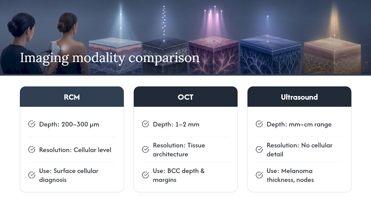
High-frequency ultrasound goes deeper still, into the millimeter-to-centimeter range, and is the better tool for measuring tumor thickness before melanoma excision or assessing lymph node involvement. It sacrifices almost all cellular detail in exchange for that depth.
RCM sits at the opposite end of that spectrum: shallowest reach, highest resolution, the only one of the three that gives a genuinely cellular-level picture comparable to histology. In practice, these tools aren’t competitors so much as a stack. A clinician might use RCM to characterize a lesion’s cellular architecture at the surface, then reach for OCT or ultrasound if depth of invasion becomes the pressing clinical question, particularly before a surgical plan gets finalized.
Where Confocal Skin Imaging Is Headed Next
The clearest trend is convergence: manufacturers are working on combining RCM with OCT in a single device, letting one exam capture both cellular detail near the surface and structural depth information below it, instead of scheduling two separate appointments. That would meaningfully shrink the current gap between RCM’s shallow reach and the clinical need to know how deep a lesion goes.
Artificial intelligence support for mosaic interpretation is the other major front. Pattern-recognition algorithms trained on large RCM image sets are being tested as a second reader, flagging pagetoid cells or tumor island silhouettes for a human reader to confirm, which could shorten the training curve that currently keeps RCM concentrated at high-volume academic centers. None of that replaces a trained reader today, but it points toward interpretation support becoming standard the way computer-aided detection already is in mammography.
Handheld probe miniaturization is also progressing, aimed at making RCM practical on genuinely difficult sites, the nail unit, mucosal surfaces, deep skin folds, where current probes struggle to maintain even contact. And ex vivo fluorescence confocal microscopy continues expanding its footprint in Mohs surgery suites, offering faster intraoperative margin reads than traditional frozen section processing, a shift that changes surgical workflow more than it changes diagnostic accuracy.
Editorial Take: What Actually Matters With RCM
The conventional pitch for RCM oversells it as a biopsy replacement, and that framing sets clinicians up to be disappointed. Read the diagnostic numbers honestly: 93.6% sensitivity and 82.7% specificity are strong for a triage tool, not strong enough to skip tissue diagnosis on a lesion that still looks concerning after imaging.
What gets underappreciated is the biopsy-targeting function. RCM’s real edge isn’t answering “cancer or not” in isolation, it’s telling you exactly where on a lesion to put the punch, which cuts down on the sampling errors that plague blind biopsies of large or heterogeneous lesions. That’s a workflow improvement, not a diagnostic replacement, and it deserves to be framed that way to referring clinicians and patients alike.
Prioritize the interpretation training gap before the equipment purchase. A wide-probe system sitting in a practice with an undertrained reader produces false reassurance, which is arguably worse than not having the technology at all. Reading skill, not laser power, is the bottleneck.
Get Confocal Imaging and Dermatopathology Support From EIV Diagnostics
Reading an RCM mosaic well takes volume and mentored feedback, and not every practice has either in-house. EIV Diagnostics gives referring clinicians a direct path to board-certified pathologists for dermatopathology correlation, without the wait times that come with sending cases to a large hospital-system lab.

That matters most in the exact moment this article has been describing: a mosaic that looks ambiguous, a biopsy that needs precise site targeting, or a case where tissue diagnosis has to confirm what imaging suggested. EIV Diagnostics’ dermatopathology services are built for that handoff, pairing confocal findings with histopathologic expertise instead of leaving clinicians to interpret borderline cases alone. For practices that also need broader diagnostic support on a case, molecular pathology testing and digital pathology review are available through the same lab. If a lesion warranted RCM in the first place, it deserves a fast, expert tissue correlation when the imaging isn’t enough on its own. Reach out to EIV Diagnostics to set up a consultation or discuss case referral for your next equivocal lesion.
Sources
- Introduction to reflectance confocal microscopy and its use in clinical practice — PMC
- Reflectance confocal microscopy — DermNet NZ
- Clinical Applications of In Vivo and Ex Vivo Confocal Microscopy — MDPI
This article is general information, not a substitute for advice from a qualified doctor. Consult a qualified healthcare professional about your own circumstances before acting on anything here.
FAQ
What is confocal microscopy in dermatology?
It’s a noninvasive imaging technique that uses a low-power laser to produce near-histologic, cellular-level images of skin, helping clinicians evaluate suspicious lesions without an immediate biopsy.
What are the downsides of using a confocal microscope on skin?
Its imaging depth is limited to about 200 to 300 micrometers, so it can’t assess deeper dermal involvement, and thick, scaly, or hyperkeratotic lesions block the laser before it reaches useful depth.
Can confocal microscopy be used to detect skin cancer?
Yes.
What is the purpose of confocal microscopy in skin evaluation?
Its main purpose is to triage equivocal lesions after dermoscopy, guide precise biopsy site selection, and monitor treatment response, reducing unnecessary excisions on lesions that turn out to be benign.
What findings show up on a normal versus abnormal RCM exam?
A normal exam shows uniform honeycomb keratinocyte patterns and edged dermal papillae, while abnormal findings include pagetoid melanocytes, disrupted architecture, or dark tumor island silhouettes that prompt biopsy, a distinction pathologists at labs like EIV Diagnostics confirm through subsequent tissue correlation.