
By EIV Diagnostics · September 20, 2026
Decode Your Melanoma Pathology Report: Breslow & Ulceration
How Breslow thickness and ulceration in your melanoma pathology report set AJCC stage, guide SLN biopsy, and when to get a specialist opinion.

A melanoma pathology report is the lab document that confirms your diagnosis and describes the tumor’s biology in detail your doctor needs for staging. Two findings drive most of the decision-making that follows: Breslow thickness, which measures how deep the tumor grew, and ulceration status, which tracks whether the skin over the tumor broke down. Read the report once for familiarity, then review it line by line with your treating clinician, who will translate it into a staging category and a treatment plan.
TL;DR:
- Breslow thickness near or above 0.8 mm or ulceration, especially combined, increases the likelihood of recommending sentinel lymph node biopsy.
- Margin status, especially if positive, often necessitates additional excision before further staging or treatment decisions.
- Regression and TIL grading are nuanced; extensive regression may underestimate tumor thickness and influence closer follow-up.
- Molecular and immunohistochemistry tests are used selectively in challenging cases or for metastasis, not routinely.
- Early-stage tumors under 0.8 mm without ulceration generally have a lower risk, but thickness and ulceration remain critical for staging and management.
Table of Contents
- What Is in a Melanoma Pathology Report?
- How Report Findings Map to Staging and Treatment
- Common Terms Patients See and What They Really Mean
- Questions to Bring to Your Clinician About the Pathology Report
- Additional Tests You May See and Why They Matter
- Turnaround Times and What Happens Next
- How Specialty Labs Support Clearer, Faster Reporting
- Explanation of Clark Level and Its Relevance in Melanoma Pathology Reports
- Interpretation of Tumor Regression and Its Impact on Prognosis
- Sentinel Lymph Node Biopsy Status and the Pathology Report
- A Clinician’s Quick Perspective: Why Reading the Report Together Matters
- Get Dermatopathology, Molecular Testing, or a Mobile Blood Draw
- Sources
- FAQ
What Is in a Melanoma Pathology Report?
Your report opens with patient identifiers and specimen information, including whether the tissue came from a biopsy (a sample) or an excision (removal of the full lesion with margins). That distinction matters because a biopsy report often triggers a second, larger surgery, while an excision report may represent your final surgical pathology.
Below that sits the final diagnosis line, naming the melanoma subtype. This is where you will see wording like superficial spreading, nodular, lentigo maligna, acral lentiginous, or desmoplastic melanoma. Subtype affects behavior. Nodular melanomas, for instance, tend to grow deeper faster than superficial spreading types, which is part of why thickness matters so much regardless of subtype.
The core prognostic fields usually appear together, often in a synoptic checklist format:
- Breslow thickness, recorded in millimeters, measuring the tumor from the top of the skin’s granular layer to its deepest point.
- Ulceration, noted as present or absent, describing whether the epidermis over the tumor has broken down.
- Margins, both peripheral (side) and deep, marked as involved or clear, sometimes with a distance in millimeters.
- Mitotic rate, the count of dividing tumor cells per square millimeter.
- Microsatellites, lymphovascular invasion, and tumor-infiltrating lymphocytes (TILs), when present or assessed.
A pathology report reading guide can help you locate each field on the printed page before your appointment, since layouts vary by lab.
How Report Findings Map to Staging and Treatment
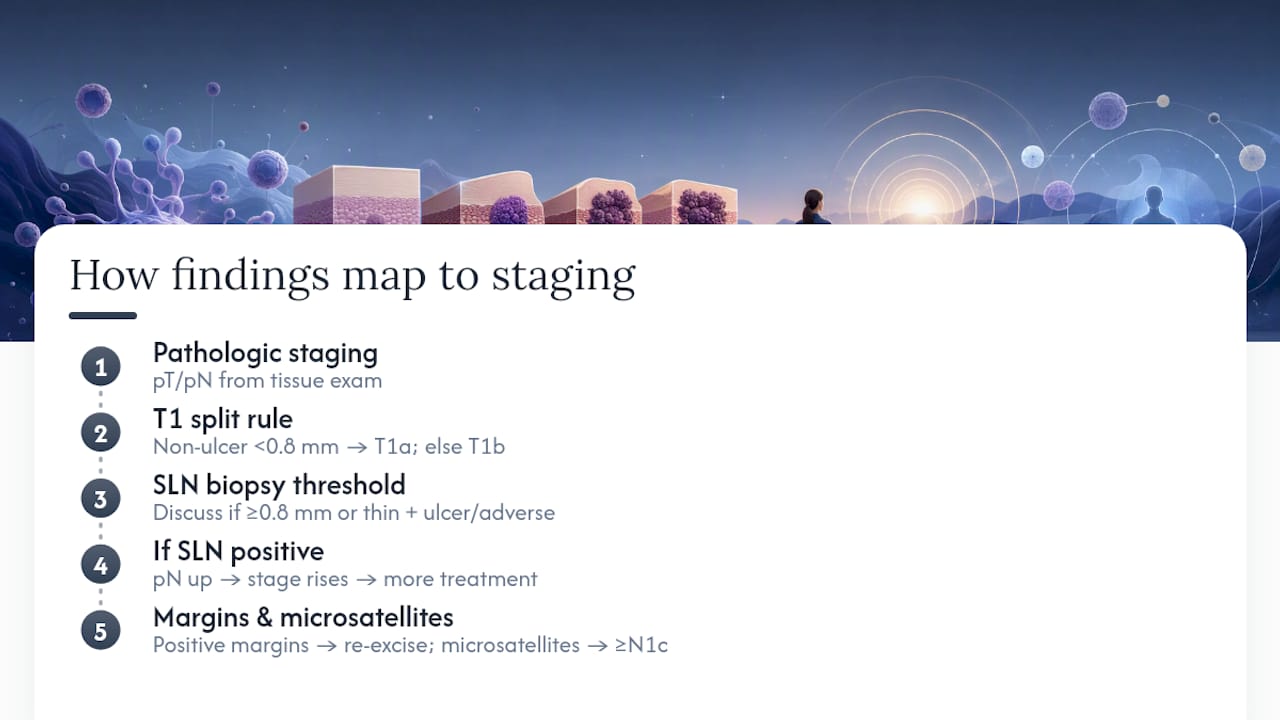
Pathologic staging uses the letter “p” before T, N, or M to signal the classification came from tissue examination rather than imaging or physical exam alone. Your pT category is built almost entirely from Breslow thickness and ulceration status.
The AJCC Eighth Edition set 0.8 mm as the dividing line within T1 tumors: a non-ulcerated melanoma under 0.8 mm is generally T1a, while one at or above 0.8 mm (or any thickness with ulceration) shifts to T1b. Pathologists round thickness to the nearest 0.1 mm, so a report reading “0.75 mm” and one reading “0.8 mm” can sit on opposite sides of that line. Ulceration alone can bump a tumor into a higher substage even when thickness stays constant, because an ulcerated melanoma has already shown it can breach the skin barrier.
Thickness and ulceration together largely determine whether your surgeon recommends a sentinel lymph node (SLN) biopsy. Tumors at or above roughly 0.8 mm, or thinner ulcerated tumors with adverse features, commonly meet the threshold for discussing SLN biopsy. If a sentinel node comes back positive, the N category changes and your overall stage rises, which can open the door to additional treatment. Positive margins usually mean a second excision is needed before any further staging conversation matters, since incomplete removal leaves residual tumor whose true depth is unknown. Microsatellites, small tumor deposits near the primary site, automatically classify a case as at least N1c and often push toward more aggressive treatment planning.
Common Terms Patients See and What They Really Mean
Some words on your report sound alarming but describe routine findings your pathologist is simply documenting, not warning you about.
- Breslow thickness: how many millimeters deep the melanoma cells reached, the single strongest predictor of behavior.
- Ulceration: loss of the skin surface over the tumor, confirmed under the microscope rather than by appearance alone.
- Mitotic rate: how many tumor cells were actively dividing in a defined area, an activity marker rather than a spread marker.
- Margins: the border of normal tissue around the tumor; “positive” or “involved” means tumor cells reach the cut edge.
- Regression: areas where the immune system appears to have already destroyed part of the tumor, which can make the remaining tumor look deceptively small.
- Tumor-infiltrating lymphocytes (TILs): immune cells found within the tumor, graded brisk or non-brisk, reflecting immune engagement rather than a simple good/bad score.
- Microsatellites: separate small tumor nests near the main lesion, distinct from satellite metastases seen clinically.
Regression and TIL grading are more nuanced than they sound. A “non-brisk” TIL result is not automatically bad news, and regression does not always mean a worse outcome. It can occasionally reflect a smaller residual tumor than the original lesion suggested.
Tissue handling can also create confusion. Epidermis sometimes tears away during specimen processing, and a pathologist may need to distinguish that artifact from true ulceration. If your report notes ulceration, it is reasonable to ask whether it was confirmed as a genuine clinical finding.
Pro Tip: Ask whether your report is written in synoptic format (a structured checklist of data elements) or narrative prose. Synoptic reports are easier to scan and less likely to omit a required field, which is why most accredited labs use them for melanoma.
Questions to Bring to Your Clinician About the Pathology Report
Walk into your follow-up appointment with a short, specific list instead of trying to remember everything on the spot.
- What is my pathologic stage, and what does it mean for my next steps?
- What is my exact Breslow thickness, and was it rounded per AJCC convention?
- Are my margins clear, and if not, what is the plan for re-excision?
- Is ulceration present, and how does that affect my staging?
- Am I a candidate for sentinel lymph node biopsy, and why or why not?
- Do any findings warrant IHC stains, molecular testing, or a second dermatopathology opinion?
Write down the answers. You will likely repeat this conversation with a surgeon, an oncologist, or both.
Additional Tests You May See and Why They Matter
Most melanoma diagnoses do not need extra testing beyond routine microscopy. But roughly 15% of pigmented lesions remain genuinely difficult to classify, and that’s when pathologists reach for confirmatory tools.
Immunohistochemistry (IHC) stains, such as SOX10 or Melan-A, help confirm melanocytic origin when a tumor looks ambiguous under standard staining. Gene expression profiling (GEP) or molecular testing may get ordered for borderline lesions, though clinicians still debate how much these results should influence treatment for early-stage disease. For metastatic or advanced melanoma, comprehensive genomic profiling of tumor tissue guides targeted therapy selection, and circulating tumor DNA (ctDNA) testing is increasingly used to monitor for recurrence after treatment.
Turnaround Times and What Happens Next
Routine melanoma pathology typically takes a matter of days, though complex cases needing special stains or outside consultation can take longer. Expect your reported stage to shift after a wide excision or sentinel lymph node biopsy, since those procedures often reveal information the original biopsy couldn’t capture. Common next steps include a surgical consult, SLN biopsy scheduling, or a dermatology referral. If your diagnosis feels uncertain or subtype-dependent, a second pathology opinion from a dermatopathologist is a reasonable and common request.
How Specialty Labs Support Clearer, Faster Reporting
Dermatopathology and molecular pathology exist specifically to reduce the ambiguity that borderline melanoma cases create. A dermatopathologist has subspecialty training in skin-specific tissue interpretation, which matters when a lesion sits at the edge between benign and malignant.
EIV Diagnostics offers dermatopathology, molecular pathology, and digital pathology services, along with mobile phlebotomy for follow-up blood draws and self-pay testing for patients acting without a standing physician order. If your case involves a difficult subtype call or you want a second read before committing to a treatment path, these are the kinds of services worth asking your care team about directly.
Explanation of Clark Level and Its Relevance in Melanoma Pathology Reports
Clark level is an older staging measure that classifies melanoma by which anatomic layer of skin the tumor has invaded, from the epidermis (Level I) down through the reticular dermis and into subcutaneous fat (Level V). It predates Breslow thickness as the primary depth measurement and was once the standard method for judging invasion.
You may still see Clark level listed on some reports, particularly for very thin melanomas, but it has largely been replaced. Breslow thickness measures actual distance in millimeters, which turned out to be a more consistent and reproducible predictor of behavior than which anatomic layer the tumor reached. Two tumors that invade the same layer can have very different thicknesses depending on how thick that skin layer happens to be at that body site.
Current AJCC staging relies on Breslow thickness and ulceration, not Clark level, for T category assignment in the vast majority of cases. If your report lists a Clark level, treat it as supplementary historical context rather than the number driving your stage. Ask your clinician to confirm which measurement, Breslow or Clark, is actually informing your treatment plan, since older report templates sometimes retain the field out of habit.
Interpretation of Tumor Regression and Its Impact on Prognosis
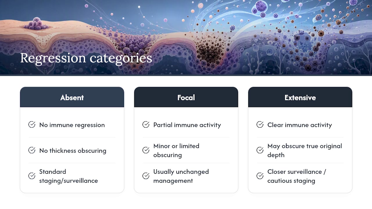
Regression describes areas within a melanoma where the immune system has already attacked and partially destroyed tumor tissue, typically visible under the microscope as fibrosis, pigment-laden immune cells, and thinned tumor nests. Your pathologist may report it as absent, focal, or extensive, sometimes with a percentage estimate.
Regression sits in genuinely nuanced territory. On one hand, it demonstrates the immune system engaged the tumor, which some clinicians view as a favorable biological sign. On the other hand, extensive regression can obscure the tumor’s true original thickness, since the deepest portion of the melanoma may have already been destroyed and replaced by scar-like tissue before the biopsy was taken. That creates a real measurement problem: your reported Breslow thickness could understate how deep the tumor actually grew.
This is why extensive regression sometimes prompts closer surveillance or a more cautious approach to staging decisions, even when the measured thickness looks reassuring. It is not a finding to interpret in isolation. Ask your clinician directly whether regression was noted on your report and whether it affected confidence in your Breslow measurement. That single question can clarify whether your stage carries more uncertainty than the printed numbers suggest.
Sentinel Lymph Node Biopsy Status and the Pathology Report
Sentinel lymph node (SLN) biopsy is a separate procedure from your original skin biopsy, performed to check whether melanoma cells have reached the first lymph node draining the tumor site. Its result becomes a second pathology report that gets merged with your original skin findings to complete staging.
A negative SLN biopsy means no tumor cells were found in the sampled node, and your N category stays at N0. A positive result means tumor cells were detected, which shifts you into an N1, N2, or N3 category depending on how many nodes are involved and whether the deposits are microscopic or clinically detectable. That shift can move you from Stage I or II into Stage III, materially changing the treatment conversation and often opening eligibility for adjuvant therapy.
Not every melanoma patient needs an SLN biopsy. The decision hinges heavily on the Breslow thickness and ulceration status from your original report, which is why clinicians wait for the full pathology before deciding whether to recommend the procedure. If you have already had an SLN biopsy, ask specifically how many nodes were removed, how many were positive, and whether the tumor deposit size was microscopic or larger, since that detail also factors into staging and treatment intensity.
A Clinician’s Quick Perspective: Why Reading the Report Together Matters
A pathology report is one input into your care plan, not the whole plan. Numbers on a page don’t capture clinical context, prior lesions, or your overall health picture, and ambiguous cases genuinely benefit from a dermatopathology second read. Keep a copy of every report you receive, and never hesitate to ask a pathologist’s office to clarify a term you don’t recognize.
— EIV Diagnostics
Get Dermatopathology, Molecular Testing, or a Mobile Blood Draw
EIV Diagnostics gives patients and clinicians a direct route to specialist pathology without routing every question through a large hospital system. If your report raised questions your original lab didn’t fully answer, that access matters more than most people realize until they need it.

EIV Diagnostics runs dermatopathology for second opinions and difficult subtype calls, molecular pathology for cases where a tumor’s genetics inform treatment, and digital pathology for sharing slide images with your broader care team. For patients who need blood work done without a trip to a draw site, mobile phlebotomy starts at $65 per visit, bringing a phlebotomist to your home or office. Patients without a standing physician order can also arrange self-pay testing directly. Whatever service you use, coordinate results with your treating clinician so testing and interpretation stay connected to your overall treatment plan. Visit the EIV Diagnostics services page to request an appointment or ask which option fits your situation.
Sources
- Melanoma Pathology - StatPearls (NCBI Bookshelf)
- Melanoma staging: Evidence-based changes in the AJCC Eighth Edition (PMC)
- Understanding Your Melanoma Diagnosis and Pathology Report — Melanoma Research Alliance
This article is general information, not a substitute for advice from a qualified doctor. Consult a qualified healthcare professional about your own circumstances before acting on anything here.
FAQ
How Do You Read a Melanoma Pathology Report?
Start with the final diagnosis line to confirm melanoma subtype, then find Breslow thickness, ulceration status, and margin status, since those three fields drive staging discussions. Bring the report to your clinician appointment and ask them to walk through each field with you, especially the pathologic stage and what it changes about your treatment plan.
What Lab Values Indicate Melanoma?
There is no single blood test value that diagnoses melanoma; diagnosis comes from tissue examination under a microscope, not lab bloodwork. The pathology report confirms melanoma through cell appearance and, when needed, immunohistochemistry stains like SOX10 or Melan-A that confirm melanocytic origin.
Is a Pathology Report the Same as a Biopsy?
No. A biopsy is the procedure that removes a tissue sample, while the pathology report is the document describing what the pathologist found after examining that sample under a microscope. One is the action; the other is the result.
What Does Melanoma Look Like Under the Microscope?
Pathologists look for atypical melanocytes with irregular shapes, abnormal growth patterns within the skin layers, and often increased mitotic activity compared to normal moles. The specific pattern, along with thickness and ulceration, helps determine the melanoma subtype recorded in your diagnosis report.
What Does It Mean if My Margins Are Positive?
A positive or involved margin means melanoma cells were found at the edge of the removed tissue, meaning some tumor may remain in the body. This typically prompts a second excision to achieve clear margins before staging and treatment planning move forward.