
By EIV Diagnostics · September 22, 2026
Telepathology for Clinicians: 4 Modalities and When to Use Each
Match the four telepathology modalities to real clinical cases—frozen sections, cytology, biopsies. Read accuracy data, rollout essentials, and EIV...
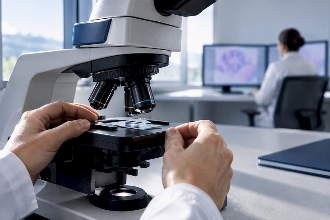
Telepathology is the practice of diagnosing tissue and cell samples remotely by sending digital pathology images over telecommunication networks instead of examining a physical slide under a microscope in person. Pathologists use it for primary diagnosis, intraoperative frozen section consults, second opinions, quality assurance, and training. The technology behind it ranges from simple photo transmission to full whole-slide imaging systems, and the modality a lab picks depends entirely on the clinical job at hand.
TL;DR:
- Telepathology offers high diagnostic accuracy, with studies showing concordance rates above 0.92 sensitivity and 0.99 specificity for specific tasks.
- Its main limitations include high setup costs, substantial storage requirements, and the need to retain physical slides for certain cases, especially in cytology.
- Successful implementation requires thorough validation, IR integration, peak load planning, and ongoing staff training to avoid workflow disruptions.
- FDA-cleared whole-slide imaging systems enable primary diagnosis, but around 5 to 10 percent of cases still require physical slides for special stains or polarized light.
- Future trends involve AI-assisted triage and cloud-based platforms that expand access across multiple sites and reduce infrastructure costs.
Table of Contents
- Overview and Brief History of Telepathology
- Telepathology Modalities Explained: Static, Dynamic, Robotic, and WSI
- How Telepathology Systems Work: Components and Workflow
- Clinical Applications and Concrete Use Cases
- What the Evidence Says About Accuracy and Turnaround
- Benefits, Limitations, and Why Adoption Isn’t Instant
- Implementation Essentials: Validation, IT, and Training
- Regulatory Status and Professional Standards
- Where Telepathology Is Headed Next
- How EIV Diagnostics Approaches Telepathology
- Get Digital Pathology Support From EIV Diagnostics
- Sources
- FAQ
Overview and Brief History of Telepathology
The term “telepathology” dates back to 1986, coined during early experiments transmitting video microscopy images over telephone and television links so a pathologist in one location could interpret slides sitting in another. Those first systems were clumsy by today’s standards: low-resolution video feeds, narrow bandwidth, and a pathologist squinting at a grainy monitor trying to make a call on a frozen section. What started as a workaround for staffing gaps in remote hospitals has become a mainstream diagnostic tool.
Telepathology is often used interchangeably with “digital pathology,” but the two aren’t the same thing. Digital pathology is the broader field, covering everything from image analysis software to AI-driven quantification. Telepathology is specifically the remote-interpretation piece of that field: the act of a pathologist rendering a diagnosis on a sample they never physically touched, using images sent to them digitally.
The evolution since 1986 tracks pretty closely with internet infrastructure itself:
- Early static image transmission over dial-up and ISDN lines in the late 1980s and 1990s
- Real-time video microscopy consultations became viable as bandwidth improved through the 2000s
- Robotic telemicroscopy let a remote pathologist actually drive the microscope stage
- Whole-slide imaging (WSI) scanners, which digitize an entire glass slide into a single high-resolution file, took over as the dominant approach once storage and network speeds caught up
Each of those stages still exists in some form today, and that’s the part worth understanding before diving into hardware specs. The right modality depends on what you’re trying to accomplish, not which one is newest.
Telepathology Modalities Explained: Static, Dynamic, Robotic, and WSI
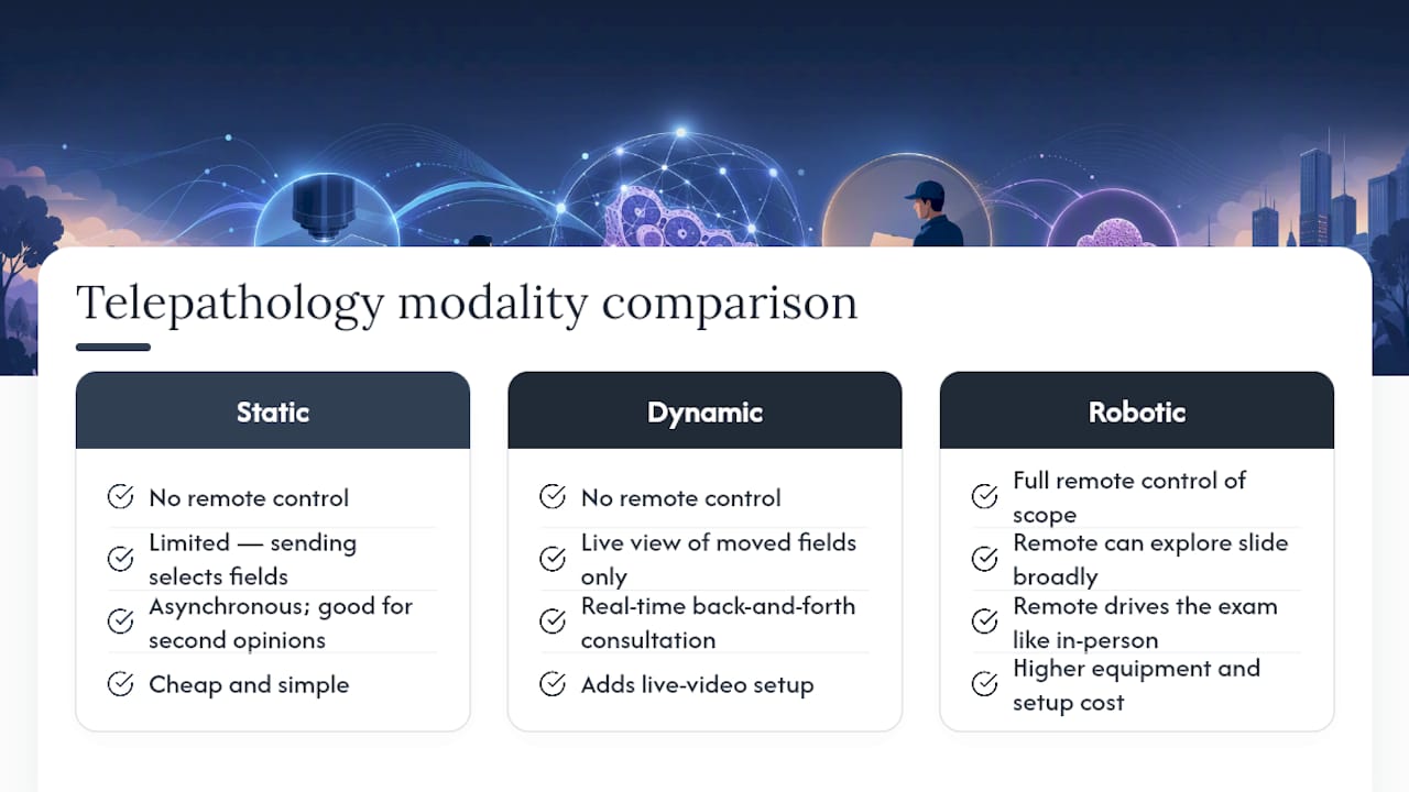
Four modalities cover nearly everything a lab or clinic will encounter, and each one trades cost against flexibility in a different way.
Static, or store-and-forward, telepathology works by capturing a fixed set of still images from a slide and sending them to a remote pathologist for review. It’s cheap and simple, requiring nothing more than a camera-equipped microscope and an email or file-sharing setup. The catch is sampling: the sending pathologist chooses which fields to capture, so anything outside those frames is invisible to the reviewer. That makes static imaging fine for straightforward second opinions but risky for complex specimens where the diagnosis hinges on a region nobody thought to photograph.
Dynamic, nonrobotic telepathology adds a live video feed, letting a remote consultant watch the microscope image in real time while the local technician moves the slide. It supports genuine back-and-forth consultation, useful for a tricky frozen section during surgery, but the remote party has no control over what they’re seeing.
Robotic telemicroscopy solves that control problem. The remote pathologist operates the microscope stage, focus, and objective lenses directly, effectively driving the exam from anywhere with a network connection. It closes the gap between remote and in-person review almost completely, but the equipment and setup cost more than either static or nonrobotic systems.
Whole-slide imaging has become the modality of choice for primary diagnosis in many settings. A scanner digitizes the entire slide at high resolution, producing a virtual slide the pathologist can pan and zoom exactly like a physical one under a scope, at any magnification, at any time. WSI eliminates the sampling bias of static imaging, but it demands serious storage and bandwidth. A single scanned slide can run into gigabytes, and a busy lab generates thousands of them.
How Telepathology Systems Work: Components and Workflow
A working telepathology setup is really four pieces stacked on top of each other: acquisition, software, transmission, and integration.
- Image acquisition. This starts with a WSI scanner for full digitization, a mounted camera for targeted still or video capture, or a simple gross-imaging camera for specimen photos before sectioning. The choice depends on volume and use case, not just budget.
- Image management and viewer software. Once a slide is digitized, someone needs to view, annotate, and route it. Viewer platforms handle zooming, measurement tools, and case assignment, and increasingly plug into AI modules for pre-screening or quantification tasks.
- Integration with the laboratory information system (LIS) and electronic medical record (EMR). This is the step labs underestimate. Without it, digital images sit disconnected from patient history, prior results, and the report itself, which defeats the point of digitizing anything.
- Transmission and storage. Large WSI files need real bandwidth and redundant storage, not a shared drive someone set up five years ago. Cloud storage has largely replaced on-premises servers for labs handling meaningful volume.
- Workflow and roles. A technician or histotechnologist typically handles acquisition, an IT or informatics team manages the pipeline, and the pathologist reviews and signs out the case, often through a queue that prioritizes urgent specimens ahead of routine ones.
Each link in that chain has to hold up under real clinical pressure. A scanner that produces gorgeous images is useless if the viewer software can’t talk to the LIS, and a fast network doesn’t help if storage runs out mid-quarter.
Clinical Applications and Concrete Use Cases
Telepathology shows up anywhere a diagnosis needs to happen without the specimen and the specialist in the same room.
- Primary diagnosis: A community hospital without an on-site subspecialist scans a biopsy and sends it to a remote dermatopathologist or hematopathologist for the definitive read, using WSI to preserve full-slide detail.
- Intraoperative frozen sections: A surgeon needs a margin status answer in minutes while a patient is still under anesthesia. Dynamic or robotic telemicroscopy fits here because it supports live interaction, letting the remote pathologist ask the technician to reposition the slide on the spot.
- Second opinions and subspecialty consults: A general pathologist flags an ambiguous case and routes it to a subspecialist elsewhere, often through the same static or WSI pipeline used for primary reads.
- Tumor boards: Multiple specialists review the same digital slide simultaneously during a case conference, something a single glass slide passed around a room simply can’t support.
- Quality assurance: Programs use stored digital slides for retrospective peer review, catching discordant reads without pulling physical slides out of storage.
- Education and training: Residents and students review annotated digital cases remotely, building a case library that doesn’t degrade the way glass slides eventually do.
A province-wide telepathology network illustrates the scale this can reach: centralizing pathology expertise across a wide geographic area improved access in areas that previously had no local subspecialist coverage, while keeping turnaround times comparable to traditional on-site review. That’s the practical argument for telepathology in one sentence: it moves the expert to the case instead of moving the case to the expert.
What the Evidence Says About Accuracy and Turnaround
The core question anyone evaluating telepathology asks is simple: does it produce the same diagnosis a pathologist would reach looking through an actual microscope? The research says yes, most of the time, with some caveats worth taking seriously.
By the numbers: Reviews of telepathology practice report high concordance between remote digital review and conventional light microscopy across a range of specimen types, and some frozen-section studies cite sensitivity and specificity as high as 0.92 and 0.99 for specific diagnostic tasks.
Those figures aren’t universal across every tissue type and every modality, and concordance tends to run lower for cases involving subtle cytologic detail or borderline morphology, the kind of judgment calls that trip up any two pathologists reviewing the same glass slide independently. Discordance in telepathology usually traces back to image quality, inadequate sampling in static systems, or unfamiliarity with a viewer’s zoom and navigation controls rather than any fundamental limitation of remote review itself.
On the operational side, the benefit that shows up most consistently is access: smaller facilities gain a route to subspecialty expertise they couldn’t otherwise staff, and urgent cases move faster when a queue-based digital workflow replaces physically shipping a slide across town.
Benefits, Limitations, and Why Adoption Isn’t Instant
Telepathology’s upside is real, but so are the friction points that keep some labs on the sidelines longer than expected.
The advantages tend to cluster around access and flexibility. A digital slide can be reviewed by multiple observers at once, archived indefinitely without degrading, and fed into AI-assisted quantification tools that a physical slide simply can’t support. That archival quality also means a case can be revisited months later for research or QA without pulling anything out of cold storage.
The limitations are just as concrete:
- Upfront cost for scanners, viewer software, and storage infrastructure runs high, and ongoing IT maintenance adds to it.
- Cytology cases sometimes need z-stacking, which is capturing multiple focal planes to compensate for cells sitting at different depths on the slide, and that adds scan time and file size.
- A meaningful minority of cases, estimated around 5 to 10 percent, still need the actual glass slide reviewed under polarized light or for certain special stains that digital scans don’t reliably capture.
- Proving return on investment to hospital administrators before they’ll fund a scanner and storage buildout is often the hardest barrier of all, harder than any technical hurdle.
Pro Tip: Keep a documented protocol for retrieving the original glass slide on short notice. Even a fully digital workflow needs a fallback for that small percentage of cases where polarization or a special stain makes the physical slide necessary.
Implementation Essentials: Validation, IT, and Training
Rolling out telepathology isn’t a hardware purchase. It’s a workflow change, and treating it that way from day one saves a lab from months of frustration later.
- Validate locally before going live. Professional guidance calls for a structured validation process, reviewing a defined sample of cases digitally and comparing results against the original glass-slide diagnosis before trusting the system for routine sign-out.
- Plan IT infrastructure around peak load, not average load. Bandwidth and storage requirements spike during high-volume scanning periods, and redundancy matters more than raw speed when a network hiccup can stall an intraoperative consult.
- Start with a triage model rather than a full switch. Route urgent and subspecialty cases through the telepathology queue first, keeping routine local sign-out running in parallel until staff trust the new system and validation is complete.
- Train continuously, not once. Pathologists and technicians both need time to adjust to viewer software navigation, and that learning curve directly affects the discordance numbers a program will see in its first months.
- Document data handling and access controls. Digital slides carry patient information just like any medical record, so storage and transmission need the same security discipline as the rest of a lab’s IT systems.
Labs that treat this as an IT project alone, buying a scanner and assuming the rest will follow, tend to stall. The ones that succeed redesign the workflow first and layer the technology on top of it.
Regulatory Status and Professional Standards
The FDA has cleared specific whole-slide imaging systems for primary diagnosis in surgical pathology, most notably the Philips IntelliSite Digital Pathology Solution in 2017 and the Leica Aperio AT2 DX in 2020. Those clearances matter because they establish that digital slide review, done on approved hardware with proper validation, meets the bar for clinical primary diagnosis rather than just secondary consultation.
Professional bodies fill in the practical detail the approvals don’t cover. The American Telemedicine Association has published modality definitions and use-case guidance. The College of American Pathologists sets accreditation-linked expectations around validation and documentation. The Royal College of Pathologists’ telepathology guidance focuses heavily on scope limits and local validation requirements before a lab relies on remote review for routine sign-out.
One regulatory nuance worth flagging: telecytology, the remote review of cytology specimens, carries its own caveats in many jurisdictions, and glass slide retention requirements often persist even in fully digital labs. Regulatory clearance for a scanner doesn’t remove the practical need to keep the physical specimen accessible.
Where Telepathology Is Headed Next
AI-assisted triage is the trend worth watching most closely. Algorithms trained to flag suspicious regions on a whole-slide image can pre-sort cases before a pathologist ever opens the file, and quantification tools built directly into WSI viewers are starting to handle repetitive counting tasks that used to eat up review time.
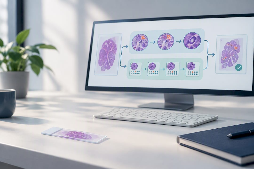
Cloud-native viewer platforms are also replacing the on-premises server model, letting labs centralize diagnostic expertise across multiple sites without each location maintaining its own storage infrastructure. Expect broader adoption as scanner costs continue to drop and validation standards mature into something closer to a shared industry playbook rather than every lab reinventing its own process.
How EIV Diagnostics Approaches Telepathology
The lab employs board-certified pathologists across various specialties and follows a validation discipline that includes documented case review before any digital workflow goes live, ensuring turnaround times appropriate for clinical needs. That approach shapes how EIV supports the providers it works with, whether that means a second opinion on a complex case or ongoing digital pathology support for a clinic without in-house subspecialty coverage. Clinics exploring telepathology implementation or needing a subspecialty second opinion can reach out to EIV Diagnostics’s provider team directly.
— EIV Diagnostics
Get Digital Pathology Support From EIV Diagnostics
Standing up a telepathology workflow from scratch means scanners, viewer software, LIS integration, and a validation process before a single case can safely go live. EIV Diagnostics gives clinics and providers a faster path: board-certified pathologists already running validated digital workflows, so you get subspecialty review without building the infrastructure yourself.

EIV Diagnostics offers digital pathology services alongside molecular pathology, dermatopathology, and mobile phlebotomy that brings specimen collection directly to a patient’s home or office starting at a price point of $65 per visit. Providers get rapid turnaround and direct pathologist access; patients with a prescription, or those who want to self-pay, can also order testing directly through EIV Diagnostics’s self-pay program. Clinics seeking subspecialty second opinions or digital pathology support for routine cases can request consultations through EIV Diagnostics’ digital pathology page.
Sources
The clinical and regulatory claims in this article draw on a small set of core references worth reading directly if you’re building a program of your own. The PMC overview of telepathology covers definitions, modalities, and concordance data. The COVID-19 era review details FDA-cleared WSI systems and modality trade-offs. Implementation guidance comes from a study on deployment factors, while limitations around glass-slide necessity are documented in a review of digital pathology’s constraints. The RCPath guidance document rounds out the validation and scope recommendations referenced throughout.
This article is general information, not a substitute for advice from a qualified doctor. Consult a qualified healthcare professional about your own circumstances before acting on anything here.
FAQ
What Is the Standard Definition of Telepathology?
Telepathology is the remote practice of pathology using digital or analog images transmitted over a network so a pathologist can render a diagnosis without physically handling the specimen. It covers primary diagnosis, intraoperative consults, second opinions, quality assurance, education, and research.
What Are the Four Main Types of Pathology Examined via Telepathology?
Telepathology commonly applies across surgical pathology, cytology, hematopathology, and dermatopathology, with the modality chosen (static, dynamic, robotic, or WSI) depending on the specimen type and urgency. Surgical and dermatopathology cases lean heavily on whole-slide imaging for primary diagnosis, while frozen sections often use dynamic or robotic setups for real-time interaction.
What Are the Main Downsides of Digital Pathology?
The biggest downsides are cost, storage demands, and the fact that roughly 5 to 10 percent of cases still require the original glass slide, particularly for polarized-light assessment or certain special stains. Cytology cases also often need z-stacking, which increases scan time and file size compared to standard tissue sections.
Is Digital Pathology FDA Approved?
Yes, specific whole-slide imaging systems have received FDA clearance for primary diagnosis, including the Philips IntelliSite Digital Pathology Solution and the Leica Aperio AT2 DX. That clearance applies to the specific cleared devices and validated workflows, not to digital pathology as a blanket category.
How Long Does It Take to Become a Pathology Technician Who Works With Telepathology Systems?
Pathology technician programs typically take one to two years to complete through an accredited certificate or associate degree program, with additional on-the-job training needed for digital scanning and imaging software specifically. Most labs layer telepathology-specific training on top of that base certification since scanner operation and viewer software aren’t part of standard technician coursework.