
By EIV Diagnostics · August 4, 2026
Serum vs Plasma: Pick the Right Sample Every Time
Discover the key differences between serum vs plasma and learn how to choose the right sample for reliable lab results every time.
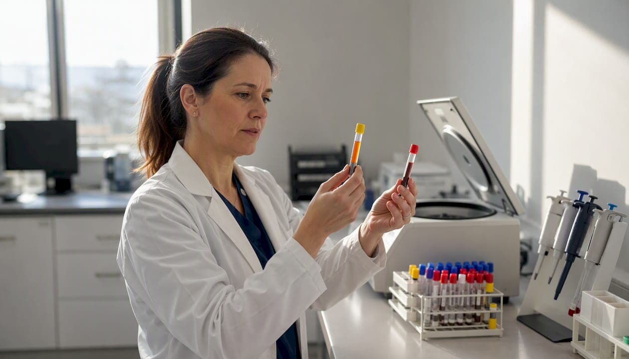
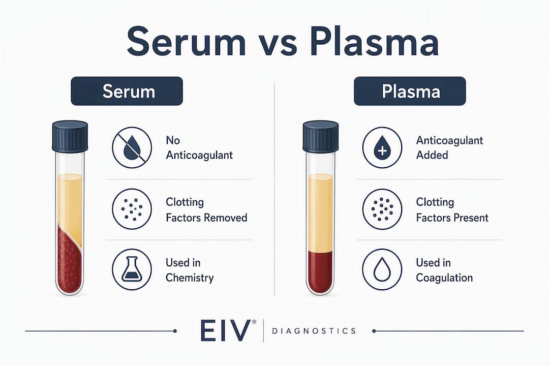
Plasma retains fibrinogen and all clotting factors; serum is what remains after those factors have done their job and the clot has been removed. That single biochemical difference determines which tube you reach for, how long you wait before centrifuging, and whether your assay result is valid.
TL;DR
- Coagulation tests (PT, aPTT, fibrinogen) require plasma collected in a citrate tube — serum cannot work because the clotting factors are already gone.
- Most routine chemistry and serology panels accept serum or plasma, but always verify with the assay manufacturer’s instructions before switching matrices.
- When speed matters, plasma wins: no clot-wait time, and heparin plasma produces more usable volume than serum from the same draw.
Table of Contents
- What serum and plasma actually contain
- How labs collect and prepare each matrix
- How anticoagulants can interfere with your assay
- Which tests prefer serum, and which prefer plasma?
- Handling, storage, and avoiding pre-analytical errors
- A quick mnemonic and decision checklist for the point of collection
- How Eivdiagnostics handles serum and plasma in practice
- Key Takeaways
- The matrix choice most labs get wrong
- Reliable specimen collection and processing at Eivdiagnostics
- Useful sources
- FAQ
What serum and plasma actually contain
Plasma accounts for roughly 55% of blood volume and is approximately 90–92% water. The remaining fraction carries proteins, electrolytes, hormones, lipids, and gases. Fibrinogen is present in normal plasma but absent in serum.
Both matrices share the major protein backbone: albumin (the dominant carrier protein), immunoglobulins like IgG, transferrin, and complement proteins. The difference is what happens when you let blood clot. Clotting consumes fibrinogen and coagulation cascade factors, converting them into a fibrin mesh that traps platelets and red cells. Centrifuge that clot away and you have serum — the same fluid minus those factors.
The clotting process is not biochemically passive. Platelets and leukocytes release proteins during clot formation that become part of serum, meaning serum sometimes measures clot-related artifacts alongside circulating analytes. One well-documented example: platelet-secreted VEGF measures roughly 230 ± 63 pg/mL in serum versus about 38 ± 8 pg/mL in plasma in normal individuals. For proteomics and biomarker discovery workflows, that difference is not a minor rounding error — it can change which proteins appear significant.
Key compositional insight: Serum is not simply “plasma without clotting factors.” The clotting event actively releases platelet-derived proteins into the matrix, so serum and plasma represent two distinct biochemical environments, not one with a subtraction.
What each matrix contains at a glance:
- Both: albumin, IgG and other immunoglobulins, electrolytes (Na⁺, K⁺, Cl⁻, HCO₃⁻), glucose, lipids, hormones, enzymes
- Plasma only: fibrinogen is present in plasma at normal concentrations, clotting factors I, II, V, VIII, XIII, von Willebrand factor
- Serum only (relatively elevated): platelet-secreted proteins (VEGF, platelet factor 4), some growth factors released during clot formation
How labs collect and prepare each matrix
Getting the matrix right starts at the tube rack, not the analyzer. The wrong tube or a missed processing step is the most common reason a specimen gets rejected before it ever reaches the instrument.
Step-by-step: serum preparation
- Collect whole blood into a plain red-top or gold-top serum separator tube (SST). SSTs contain a gel barrier that migrates between clot and serum during centrifugation.
- Allow the blood to clot undisturbed at room temperature for several minutes. Do not invert or agitate — mechanical disruption breaks the forming clot and leaves fibrin strands that clog analyzers.
- Centrifuge at moderate speed for about 10 minutes in a refrigerated centrifuge.
- Transfer the supernatant (serum) immediately into a clean labeled tube. Do not let serum sit on the clot — cellular metabolism continues and will shift potassium and glucose values within minutes.
Step-by-step: plasma preparation
- Collect into the appropriate anticoagulant tube: lavender (EDTA), green (lithium or sodium heparin), or light blue (sodium citrate) depending on the assay.
- Invert gently 8–10 times to mix anticoagulant with blood. Do not shake.
- Centrifuge promptly. Standard plasma centrifugation is at moderate speed for about 10 minutes; platelet-poor plasma may require higher speed and longer time., sometimes with a second spin.
- Pipette the plasma layer carefully, avoiding the buffy coat.
Collection variables at a glance
| Tube color | Additive | Matrix yielded | Primary uses | Centrifuge settings | Processing timing |
|---|---|---|---|---|---|
| Red (plain) | None | Serum | General chemistry, serology | Moderate speed centrifugation | After clot time |
| Gold / SST | Clot activator + gel | Serum | Routine chemistry panels | 1,000–2,000 × g × 10 min | After 15–30 min clot time |
| Lavender | EDTA | Plasma | CBC, molecular, toxicology | 1,000–2,000 × g × 10 min | Immediately after draw |
| Green | Lithium/sodium heparin | Plasma | Stat chemistry, electrolytes | 1,000–2,000 × g × 10 min | Immediately after draw |
| Light blue | Sodium citrate | Plasma | PT, aPTT, coagulation | ~2,000 × g × 15 min | Immediately; fill to line |
Pro Tip: An underfilled citrate tube is one of the most common causes of falsely prolonged PT/aPTT results. The citrate-to-blood ratio must be 1:9; if the tube is less than 90% full, reject and redraw. The same logic applies to any tube with a fixed anticoagulant volume.
How anticoagulants can interfere with your assay
Choosing plasma does not end the decision. EDTA, heparin, and citrate each interact with assay chemistry in ways that can produce meaningfully different results — or outright invalid ones.
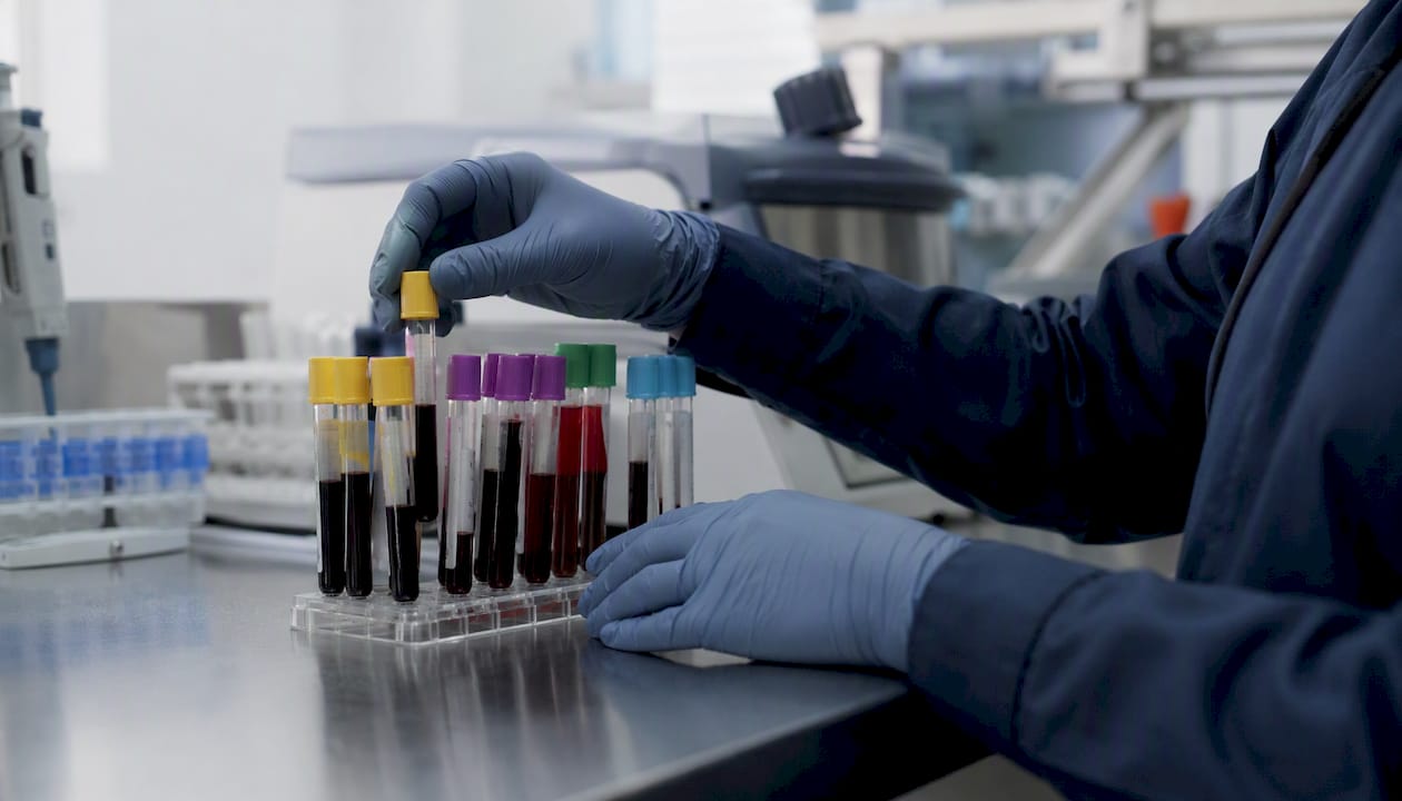
EDTA chelates divalent cations, particularly calcium and magnesium. That mechanism is exactly why it works as an anticoagulant, but it also means EDTA plasma is incompatible with calcium-dependent enzyme assays and any test that requires intact metal cofactors. Ionized calcium measurements on EDTA plasma are simply wrong. Trace-metal assays are also problematic because EDTA itself introduces metal contamination at the concentrations used in standard tubes.
Heparin works by activating antithrombin III rather than chelating cations, so it leaves calcium intact. That makes lithium heparin plasma the preferred matrix for most stat chemistry panels and electrolyte testing. The caveat: heparin can introduce endotoxin-like effects that impact downstream cellular and functional assays, and it interferes with PCR-based molecular tests by inhibiting polymerase activity. Never use heparin plasma for molecular diagnostics.
Citrate binds calcium reversibly, which is why it is the anticoagulant of choice for coagulation studies — the calcium can be added back in the analyzer to trigger clotting in a controlled way. The critical fill-volume requirement (1:9 citrate-to-blood ratio) is not a suggestion. An underfilled tube dilutes the plasma and artificially prolongs clotting times; an overfilled tube leaves excess calcium that shortens them.
- EDTA plasma: avoid for ionized calcium, calcium-dependent enzymes, trace metals, PCR
- Heparin plasma: preferred for stat chemistry and electrolytes; avoid for molecular/PCR assays and some functional cellular tests
- Citrate plasma: required for PT, aPTT, fibrinogen, D-dimer; fill to the line every time
Researchers should treat plasma as a family of three distinct matrices, not a single category. EDTA, heparin, and citrate plasma each carry method-specific interactions that can change results compared with serum or with each other.
Which tests prefer serum, and which prefer plasma?
The honest answer is: check the assay manual first. That said, strong patterns exist across test categories, and knowing them prevents most ordering errors.
Serum can yield higher apparent concentrations for some metabolites and biomarkers because the clotting event releases cellular contents into the matrix. Cardiac troponin is a classic example — serum is often the historically validated matrix, though many modern high-sensitivity assays have been validated for both. Plasma is favored when faster turnaround is required, because eliminating the 15–30 minute clot wait can meaningfully compress total lab turnaround time in high-volume or emergency settings.
| Test category | Preferred matrix | Rationale / notes |
|---|---|---|
| PT, aPTT, fibrinogen, D-dimer | Citrate plasma | Clotting factors must be intact; serum cannot be used |
| Comprehensive metabolic panel (CMP) | Serum or heparin plasma | Most analyzers validated for both; check reference ranges |
| Cardiac troponin | Serum (historically); plasma validated on many platforms | Confirm with assay insert; some platforms show matrix bias |
| Serology (ANA, HIV Ab, RPR) | Serum | Most kits validated in serum; plasma may require separate validation |
| CBC, differential | EDTA whole blood | Not a serum/plasma test — listed for completeness |
| Therapeutic drug monitoring | Serum or heparin plasma | Heparin interference varies by drug; check manufacturer guidance |
| Proteomics / biomarker discovery | Plasma (EDTA or heparin) | Lower platelet-protein contamination; more reproducible baseline |
| Molecular / PCR | EDTA plasma or whole blood | Heparin inhibits PCR; citrate acceptable for some assays |
| Toxicology | EDTA plasma or serum | Matrix varies by analyte; confirm with lab protocol |
For comprehensive metabolic panel testing, most modern analyzers accept either serum or lithium heparin plasma, but reference ranges were historically established in serum. Switching matrices mid-patient-series without revalidation can introduce a systematic bias that looks like a clinical change.
Serum and plasma are not interchangeable for all assays; switching matrices requires validation through spike-and-recovery testing, dilution linearity, and paired sample comparison before assuming equivalence.
Pro Tip: Before ordering a test in a matrix the lab has not previously used, ask for the assay’s package insert or method sheet. The manufacturer’s validated matrix is the only defensible starting point. “It probably works” is not a validation.
Handling, storage, and avoiding pre-analytical errors
Pre-analytical errors account for the majority of laboratory mistakes, and most of them happen before the sample reaches the analyzer. Hemolysis, lipemia, delayed processing, and platelet carryover are the four most common culprits.
Delayed processing and incomplete clotting affect analytes like potassium and phosphorus; hemolysis and platelet carryover introduce biases that can invalidate results entirely. A hemolyzed serum sample releases intracellular potassium, LDH, and AST into the matrix — the analyzer cannot distinguish those from circulating levels.
Storage temperature guidelines
- Process specimens within 2 hours of collection when possible. For serum, this means completing the clot step and centrifuging before the 2-hour mark.
- Store separated serum or plasma at 2–8°C for short-term holding (generally up to 8 hours for most routine analytes, shorter for labile ones like ammonia or ACTH).
- For longer storage, freeze at −20°C for most proteins and chemistry analytes. Freeze at −80°C for sensitive biomarkers, cytokines, and proteomics samples.
- Limit freeze-thaw cycles. Most analytes tolerate one to two cycles; beyond that, protein degradation and precipitation become significant. Aliquot before freezing to avoid repeated thawing of the same tube.
- After thawing, re-centrifuge at 1,000–2,000 × g for 5 minutes to pellet any cryoprecipitate before pipetting.
Common pre-analytical problems and their effects:
- Hemolysis: elevates K⁺, LDH, AST, phosphorus; interferes with spectrophotometric assays
- Lipemia: scatters light and causes turbidity interference in photometric methods; can falsely lower sodium (pseudohyponatremia) in older indirect ISE methods
- Platelet carryover: elevates VEGF, platelet factor 4, and potassium in plasma if the buffy coat is disturbed during pipetting
- Delayed processing: potassium rises, glucose falls, pH shifts — all from ongoing cellular metabolism
- Incomplete clotting (serum): fibrin strands clog analyzer probes and cause false flags or sample rejection
Pro Tip: When aliquoting frozen plasma for a multi-site study, label each aliquot with the freeze date and cycle number. A tube on its third thaw cycle that gets mixed into a first-thaw batch will introduce variability that looks like biological noise — and it will take weeks to trace.
A quick mnemonic and decision checklist for the point of collection
Memory aids work best when they are short enough to recall under pressure. Two phrases cover the core logic:
P = Preserved (Plasma: anticoagulant preserves the clotting factors) S = Separated (Serum: clotting factors are gone, separated out with the clot)
That framing holds up at the tube rack. Plasma is what you get when you prevent clotting; serum is what you get after clotting finishes. Mnemonic framing helps field teams make quicker, lower-error decisions at the point of draw, but the analyte and assay validation still drive the final call.
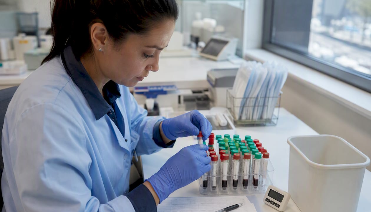
Decision checklist: serum or plasma?
Work through these questions in order:
- Is this a coagulation test (PT, aPTT, fibrinogen, D-dimer)? Yes → citrate plasma only. Stop here.
- Does the assay require intact clotting factors or is it sensitive to anticoagulant interference? Yes → serum or check assay manual for compatible anticoagulant.
- Is this a molecular/PCR-based test? Yes → EDTA plasma or whole blood. Never heparin.
- Is speed critical (stat, ED, dialysis)? Yes → heparin plasma eliminates clot-wait time and produces more volume per draw.
- Has this patient had prior results in a specific matrix? Yes → match the prior matrix to maintain longitudinal comparability.
- Does the assay manufacturer specify a matrix? Always check the package insert before assuming equivalence.
- Is this a research or proteomics workflow? Prefer EDTA or heparin plasma for lower platelet-protein background.
- None of the above applies? Default to serum (SST) for routine chemistry and serology unless the lab protocol specifies otherwise.
Document the chosen matrix in the test order. For longitudinal studies and chronic-disease monitoring, locking matrix consistency is as important as locking the assay platform — switching mid-series without revalidation introduces a systematic offset that can mimic disease progression.
How Eivdiagnostics handles serum and plasma in practice
Eivdiagnostics operates as an independent pathology laboratory in Fresno, CA, with board-certified pathologists overseeing specimen workflows across clinical pathology services that include routine chemistry, molecular diagnostics, toxicology, and specialized panels.
Every specimen type follows tube-specific centrifugation protocols aligned with the parameters described in this article: serum tubes complete the full clot step before spinning, citrate tubes are checked for fill adequacy before processing, and EDTA plasma is kept separate from heparin plasma in the workflow to prevent anticoagulant cross-contamination. Pre-analytic rejection criteria are applied at accessioning — hemolyzed, lipemic, or underfilled specimens are flagged before they reach the analyzer, not after.
For patients and providers who cannot come to the lab, Eivdiagnostics offers mobile phlebotomy, bringing a trained phlebotomist to the patient’s home or office. That service matters for matrix integrity: a specimen drawn, processed, and transported correctly from the point of collection produces far fewer re-draws than one collected elsewhere and shipped without temperature control.
Key service capabilities relevant to serum and plasma work:
- Tube-specific collection and processing protocols for serum, EDTA plasma, heparin plasma, and citrate plasma
- Board-certified pathologist review for complex or ambiguous results
- Mobile phlebotomy for home and office draws with proper cold-chain handling
- Direct-to-consumer test ordering — no physician order required for many panels
- Consults available for providers selecting the correct matrix for specialized or research assays
Eivdiagnostics works with providers, insured patients, and self-pay patients. If you are unsure which matrix your test requires, the lab’s clinical team can advise before the draw.
Key Takeaways
Plasma retains fibrinogen and all clotting factors; serum is the cell-free fluid remaining after those factors have formed and been removed with the clot, making the two matrices biochemically distinct and not universally interchangeable.
| Point | Details |
|---|---|
| Core difference | Plasma contains fibrinogen at normal concentrations and clotting factors; serum does not — they are consumed during clotting. |
| Coagulation tests always need plasma | PT, aPTT, fibrinogen, and D-dimer require citrate plasma; serum cannot substitute. |
| Plasma speeds up the workflow | Heparin plasma produces more usable fluid than serum from the same draw and eliminates the 15–30 minute clot-wait time. |
| Anticoagulant choice matters | EDTA, heparin, and citrate each interfere with different assays; always confirm compatibility with the assay manual. |
| Eivdiagnostics | Offers tube-specific processing, board-certified pathologist oversight, and mobile phlebotomy for correct specimen collection. |
The matrix choice most labs get wrong
The conventional framing of serum vs plasma as a simple binary — “serum for chemistry, plasma for coagulation” — is accurate enough to pass a board exam and wrong enough to cause real problems in practice.
The part that gets underestimated is the operational inertia around serum. Many labs built their reference ranges, their QC targets, and their longitudinal patient datasets entirely in serum. When a high-volume setting switches to plasma for efficiency reasons (and the efficiency argument is strong — faster turnaround, more volume, fewer fibrin-strand analyzer jams), those historical reference ranges no longer apply without revalidation. That revalidation step is not complicated, but it is real work, and it is frequently skipped.
The plasma adoption trend in emergency departments and dialysis centers is well-founded. Eliminating clot-wait time in a stat workflow is a genuine operational gain, and the risk of incomplete clotting causing fibrin interference in serum is a daily problem in high-throughput labs. But the right move is not to switch and assume equivalence — it is to switch with paired validation data in hand.
The other underappreciated issue is within-plasma variability. Clinicians and researchers often write “plasma” in a protocol as if it were a single matrix. EDTA plasma and heparin plasma are not the same thing for every assay. A cytokine panel validated in EDTA plasma will not necessarily perform identically in heparin plasma. Treating “plasma” as one category is how multi-site studies end up with systematic inter-site biases that take months to diagnose.
The checklist and mnemonic in this article are genuinely useful at the point of collection. But the deeper discipline is documentation: lock the matrix, lock the tube lot, and do not change either mid-study without a revalidation plan.
Reliable specimen collection and processing at Eivdiagnostics
Getting the right result starts with getting the right sample. Eivdiagnostics provides clinical pathology services built around the exact tube-specific, centrifugation-verified workflows this article describes — so the specimen that reaches the analyzer is the one the assay was validated for.

For patients who cannot travel to a lab, mobile phlebotomy brings a trained collector to your home or office, with proper cold-chain handling from draw to processing. For providers ordering specialized panels or switching matrices on an existing patient series, Eivdiagnostics’ clinical team can advise on matrix compatibility and revalidation requirements before the draw happens — not after a rejected specimen.
Eivdiagnostics accepts provider orders, patient prescriptions, and direct self-pay orders. To schedule a draw, request a consult on matrix selection, or order a panel directly, contact Eivdiagnostics or visit the clinical pathology services page to get started.
Useful sources
These are the highest-value references used in this article. Consult them for protocol specifics, centrifugation parameters, and deeper reading on anticoagulant effects.
- Physiology, Blood Plasma — StatPearls, NCBI Bookshelf: primary reference for plasma composition, volume percentages, and standard centrifugation parameters for serum and plasma preparation.
- Chemistry Testing on Plasma Versus Serum Samples in Dialysis Patients — PMC: practical clinical comparison of serum and plasma in high-volume settings; covers volume yield differences, operational efficiency, and pre-analytic error sources.
- Serum/Plasma Preparation Protocol — University of Cincinnati MMPC: step-by-step laboratory protocol for serum and plasma separation; use for centrifugation parameters and transfer timing.
- Procedure for Separating Serum and Plasma from Whole Blood — NCI Frederick: standardized SOP for biospecimen separation; relevant for research labs and biobanking workflows.
- PLOS ONE — Biomarker differences between serum and plasma: data on concentration differences for metabolites and biomarkers across matrices; useful for sensitivity trade-off discussions.
FAQ
What is found in plasma but not in serum?
Fibrinogen (normally 200–400 mg/dL) and the active coagulation cascade factors (including factors I, II, V, VIII, and XIII) are present in plasma but absent in serum, because the clotting process consumes them.
Which is better for testing, serum or plasma?
Neither is universally better. Coagulation tests require citrate plasma; many chemistry and serology panels work in either matrix. When speed matters, plasma is faster because it skips the 15–30 minute clot step and yields more volume per draw.
Why is serum used instead of plasma for some tests?
Many assay reference ranges and validation datasets were historically established in serum, so switching to plasma requires revalidation. Serum also avoids anticoagulant interference, which matters for assays sensitive to EDTA or heparin.
How do you remember the difference between plasma and serum?
Use the mnemonic: P = Preserved (plasma keeps clotting factors intact via anticoagulant) and S = Separated (serum is what remains after the clot separates out). The anticoagulant tube gives you plasma; the plain or SST tube gives you serum.
Can Eivdiagnostics help me choose the right matrix for my test?
Yes. Eivdiagnostics’ clinical team can advise on matrix compatibility and tube selection before the draw, and the lab’s mobile phlebotomy service ensures specimens are collected and handled correctly from the start.
Recommended
- Lab Test Packages | EIV Diagnostics | EIV Diagnostics
- PCR vs Rapid Test: Which COVID Test Is Right for You? | EIV Diagnostics | EIV Diagnostics
- Biospecimen & CRO Services | EIV Diagnostics | EIV Diagnostics
- Molecular Diagnostic Testing: What Patients and Clinicians Need to Know | EIV Diagnostics | EIV Diagnostics