
By EIV Diagnostics · November 8, 2026
Digital Pathology Accuracy: What the Evidence Shows
How accurate is digital pathology compared with glass slides? See concordance data, how labs validate systems under CAP and FDA rules, and what affects results.
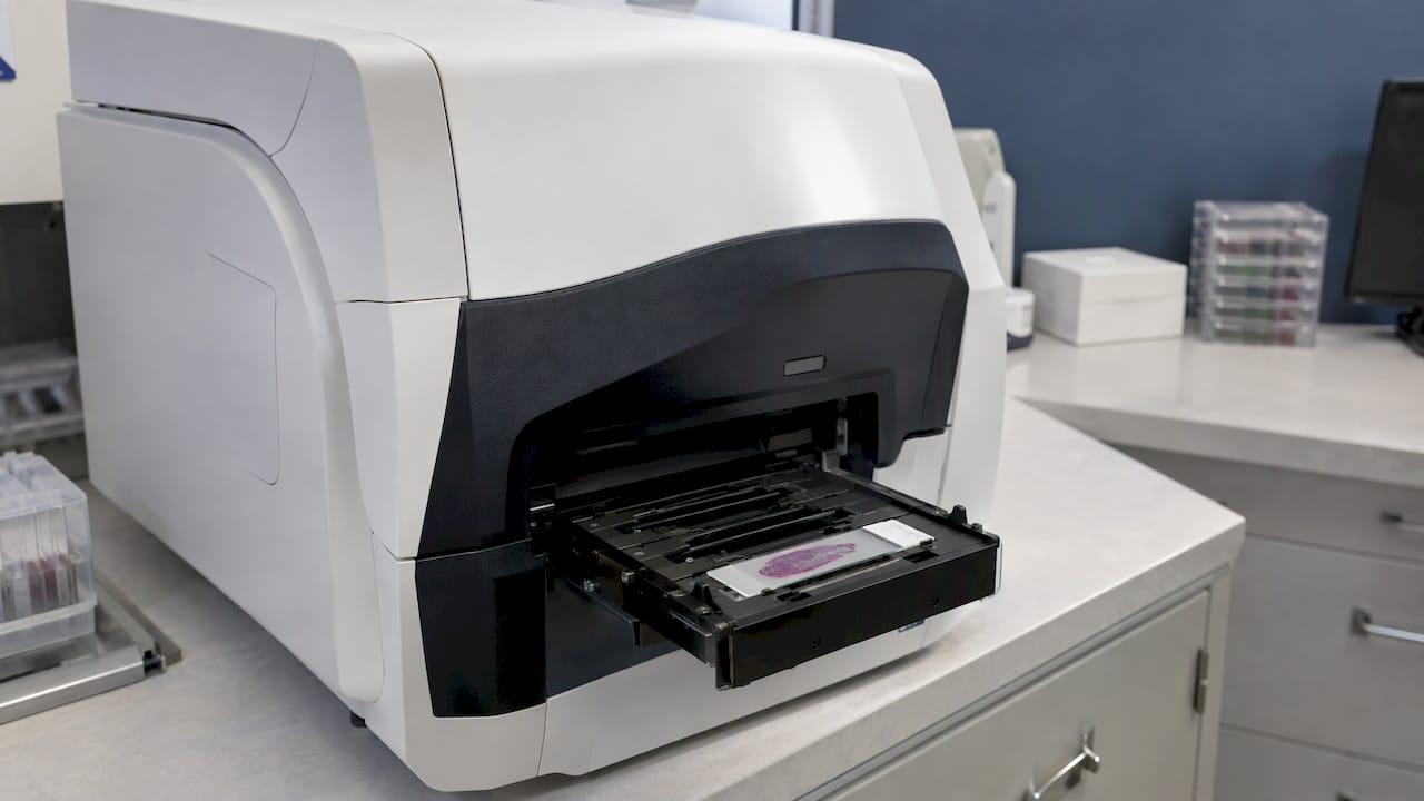
Digital pathology is as accurate as light microscopy for routine primary diagnosis when the system has been validated for its intended use. A 2021 meta-analysis found 98.3% clinical concordance between the two methods. That figure comes with conditions, though. Accuracy depends on the scanner, the slide, the case type, the pathologist and the validation behind the workflow. This guide covers what the studies measured, how labs prove a system works, and what to ask about any digital pathology service.
TL;DR
- Across 24 to 25 studies, digital reads matched glass-slide reads in 98.3% of cases for clinical concordance, and 92% for complete concordance.
- A large blinded US trial found major discordance of 4.9% for whole slide imaging and 4.6% for microscopy, a statistically noninferior result.
- The College of American Pathologists (CAP) guideline asks each lab to validate its own system with at least 60 routine cases per application, and to aim for 95% concordance.
- Accuracy is not one universal number. It depends on intended use, case complexity, image quality and ongoing monitoring.
Table of Contents
- What “accuracy” means in digital pathology
- How digital pathology compares with glass slides
- What affects accuracy in practice
- How a lab validates a digital pathology system
- The US regulatory layer
- AI-assisted reads and long-term monitoring
- Questions to ask any digital pathology provider
- Our perspective on digital pathology accuracy
- Digital pathology with EIV Diagnostics
- FAQ
- Sources
What “accuracy” means in digital pathology
People use the word loosely, so it helps to know what studies actually report. A 2021 review of digital microscopy validation lists the main measures:
- Concordance rate: how often a diagnosis made on a digital image matches the diagnosis on the glass slide.
- Kappa agreement: concordance corrected for agreement by chance.
- Accuracy against a gold standard: how often a read matches a reference diagnosis, usually a consensus or original sign-out.
- Repeatability and reproducibility: whether the same reader, or different readers and sites, reach the same answer.
Diagnostic confidence is a primary measure in that review. Diagnostic time and image quality are secondary measures.
Some studies also report sensitivity and specificity. A 2022 systematic review found that in the studies reporting good whole slide imaging (WSI) performance, sensitivity ranged from 86% to 100% and specificity from 75% to 100%. Positive predictive values ran from 92% to 99%.
One point matters for interpretation. Perfect agreement is not expected, even between two pathologists looking at the same glass slide. The review authors note that 100% concordance or a kappa of 1.0 cannot be expected from a validation study. Pathology involves judgment, and some borderline cases will always draw different calls from different readers.
How digital pathology compares with glass slides
Four studies give the clearest picture. They differ in design, so read them together rather than picking one number.
| Study | Design | Key result |
|---|---|---|
| Journal of Clinical Pathology, 2021 | Systematic review and meta-analysis of histology studies | Clinical concordance 98.3% (95% CI 97.4 to 98.9); complete concordance 92% (95% CI 87.2 to 95.1) |
| Mukhopadhyay et al., 2017 | Multicenter blinded randomized noninferiority study, 1,992 cases, 4 institutions | Major discordance with the reference diagnosis 4.9% for WSI and 4.6% for microscopy; difference 0.4% |
| Wack et al., 2016 | Multisite validation, over 10,000 pairwise comparisons | Inter-reader agreement 79.1% digital (weighted kappa 0.72) versus 76.5% glass (0.68) |
| Wharton et al., 2025 | Study of one commercial WSI system | Accuracy difference versus manual microscopy of −0.61%, meeting the noninferiority endpoint |
What the meta-analysis tells you
The 2021 meta-analysis pooled thousands of histology samples. It concluded that digital pathology performs equivalently to light microscopy for routine diagnosis. Of the major discordances it recorded, 57% related to nuclear atypia: grading of dysplasia and malignancy. These are the judgment-heavy calls where pathologists disagree with each other on glass too.
The authors also flagged limits. Most studies gave no basis for their sample size and no power calculation. And because variability on the existing microscope platform is unknown, it is not possible to say how many discrepancies come from the viewing method itself.
What the pivotal trial tells you
The 2017 study is useful because of its design: blinded, randomized and across several institutions, covering H&E, immunohistochemistry and special stains. The 0.4% difference in major discordance had a confidence interval of −0.30% to 1.01%. That supported the conclusion that WSI is noninferior to microscopy for primary diagnosis in surgical pathology.
What the reproducibility data tells you
A system can be accurate on average and still be inconsistent. The 2025 study of one FDA-regulated system measured precision as well. Agreement was 89.3% between systems and sites, 90.3% between days and 90.1% between readers, and the study met its predetermined endpoints. Precision numbers sit lower than concordance numbers because they test a harder question: whether the same slide gets the same answer across devices, days and people.

What affects accuracy in practice
The image a pathologist reads is the end of a chain. The validation review describes a digital workflow of specimen preparation, whole-slide image acquisition, slide retrieval and the viewing workstation. Each step can introduce artifacts or limits.
Before the scanner. Tissue fixation, sectioning thickness, staining quality and coverslipping all shape the image. A fold, bubble or faint stain is a problem on glass, and it is a problem on screen too. Our article on biopsy processing steps covers how to reduce recuts and overprocessing upstream.

At the scanner. Color calibration, resolution and focus matter. The scanner must also capture all of the glass material. The CAP guideline asks labs to confirm that all glass-slide material is present in the digital image.
At the workstation. Display quality, software and the reader’s training influence the read. The review also names three sources of bias in validation studies: case complexity, pathologists’ diagnostic experience and case recall.
In the case mix. A system validated on routine biopsies tells you little about a rare or difficult subspecialty. This is why validation is application-specific, according to the Digital Pathology Association (DPA). It says WSI should not be used for a clinical purpose other than the one validated unless that purpose is separately validated.
How a lab validates a digital pathology system
Validation is the step that turns published accuracy into accuracy at one specific lab. The CAP published its original WSI validation guideline in May 2013, with 12 statements. The 2021 update has three strong recommendations and nine good practice statements, and largely reaffirms the original.
The core requirements are:
- Validate the whole system, not just the scanner, in an environment that emulates the lab’s real clinical practice, with trained pathologists.
- Use at least 60 routine cases per application, with more where the application is more complex.
- Compare the same observer’s digital and glass diagnoses at least two weeks apart, to limit recall bias.
- Confirm that all glass-slide material is in the digital image.
The CAP guideline text sets these out. The 95% concordance threshold is not arbitrary. The CAP explains that the weighted mean concordance across 33 studies in its systematic review was 95.2%, which became the basis for a 95% target. It also states that 100% is the ideal but does not reflect the subjective nature of glass-slide pathology.
The DPA adds that validation matters for compliance too. It is needed for CLIA, state regulations, The Joint Commission and CAP accreditation, and to protect patient safety and data. Lab managers can find practical steps in our guides on telepathology validation for clinics and reaching 95% concordance.
Rigor in documenting quality is not unique to pathology. Other testing fields publish their own standards, such as certificate of analysis standards for lot-level lab results. The principle is the same: show your method, and tie each result to a documented process.
The US regulatory layer
Validation by the lab sits on top of FDA oversight of the device. Under 21 CFR § 864.3700, a whole slide imaging system is a Class II device subject to special controls. Premarket submissions must include component and system bench testing, precision and reproducibility data from multiple systems, sites and readers, and clinical data showing non-inferiority to an optical microscope.
The FDA authorized the first WSI device in 2017 through the de novo pathway, and cleared a second in 2019. Its 2016 guidance on technical performance assessment sets out what technical data a WSI system should have to support its intended use.
The FDA is candid about open problems. It describes two regulatory science gaps: no standardized test methods linking technical performance to clinical performance, and a lack of statistical methods for studying image quality against pathologist or algorithm performance. For readers, the practical meaning is that clinical validation, not a specification sheet, is what shows a system works.
AI-assisted reads and long-term monitoring
Accuracy for software that analyzes images is a different question from accuracy for a human reading a digital slide. An NCI workshop report notes that AI validation generally requires much larger datasets, often thousands of slides, to test robustness and reduce demographic or disease-specific bias. The 60-case rule applies to concordance of human reads, not to AI tools.
Two habits keep accuracy honest over time:
- Revalidate when something changes. A new scanner, software version or new use case calls for fresh validation, in line with the DPA principle above.
- Monitor continuously. Track concordance on a sample of cases, rescans, deferrals to glass and turnaround, and review the trends.
One caution applies to the published literature. The 2022 systematic review found high accuracy by traditional measures, but overdiagnosis was not reported in any of the studies. That is a reason for labs to keep measuring real-world outcomes after go-live, not a reason to distrust validated systems.
Questions to ask any digital pathology provider
Whether you are a clinician sending specimens or a lab manager choosing a workflow, these questions get to the substance:
- Was the system validated for the specimen types and applications I send, using the CAP approach?
- Is the reading pathologist board-certified, and does that person have subspecialty experience in my case type, such as dermatopathology?
- What happens when an image is not diagnostic quality? Is there a path back to the glass slide?
- How does the lab handle consultation and second opinions on difficult cases? Our piece on second opinions in pathology explains why review matters.
- What accreditation and inspection readiness does the lab maintain? See our overview of pathology lab accreditation.
For a broader look at when digital works well and when glass is still needed, read our post on digital pathology concordance.
Our perspective on digital pathology accuracy
We see digital pathology accuracy as the product of three things: a validated system, a well-prepared slide and a qualified pathologist. The published concordance numbers are encouraging. But a number from a study is only a starting point for the lab that signs out your case.
At EIV Diagnostics, our approach is built on board-certified pathologists with subspecialty expertise, working in a CAP-accredited laboratory. We pair digital pathology with molecular pathology and dermatopathology, so a difficult case can draw on more than one view of the same patient. Where confocal microscopy or PCR can add information, the pathologist has it.
We also believe the digital workflow is what makes rapid, consistent reporting possible for providers across the Central Valley. Images can be reviewed quickly, shared for consultation and archived for comparison. That supports timely care without giving up the standard of the diagnosis.
Our advice to providers is simple: choose a lab that can explain how its digital workflow was validated, who reads the slides and how quality is monitored. We are glad to have that conversation.
EIV Diagnostics
Digital pathology with EIV Diagnostics
EIV Diagnostics is an independent pathology laboratory in Fresno, CA. We serve providers and patients across Fresno, Clovis, Madera, Visalia and the surrounding Central Valley counties. Our digital pathology services are reviewed by board-certified pathologists, and we also offer dermatopathology, molecular pathology, histopathology and cytology.
If you are a clinician, you can send specimens for digital review through our team. If you are a patient, our self-pay testing is available with or without a doctor’s order, and mobile phlebotomy brings the blood draw to your home or office, from $65 per visit. For the full list of what we run, see all EIV Diagnostics services.
FAQ
Is digital pathology as accurate as a microscope?
For routine primary diagnosis with a validated system, yes. A 2021 meta-analysis found 98.3% clinical concordance with light microscopy, and a 1,992-case trial found WSI noninferior to microscopy. Results depend on the lab having validated the system for the case types it reads.
How many cases are needed to validate a digital pathology system?
The CAP guideline recommends at least 60 routine cases per application. The same pathologist should read each case on glass and on screen, with at least two weeks between the two reads. The lab should aim for at least 95% concordance.
Is digital pathology approved by the FDA?
Whole slide imaging systems are Class II devices under 21 CFR § 864.3700. The FDA authorized the first one in 2017 and cleared another in 2019. Each lab must still validate the system it uses for its own clinical practice.
Why do pathologists sometimes disagree, even on glass slides?
Pathology involves interpretation, especially in grading dysplasia and judging nuclear atypia. Those were the most common sources of major discordance in the 2021 meta-analysis. That is why validation targets 95% concordance rather than 100%.
Can AI make digital pathology more accurate?
AI tools may support pathologists, but they need their own validation, generally on much larger datasets than the 60 cases used for human concordance studies. Performance should be monitored over time, whatever tools a lab uses.
Sources
- Journal of Clinical Pathology: systematic review and meta-analysis of digital pathology versus light microscopy, 2021
- Mukhopadhyay et al., Whole Slide Imaging Versus Microscopy for Primary Diagnosis, 2017
- Wharton et al., Roche Digital Pathology Dx study, 2025
- Wack et al., multisite validation of whole slide imaging, 2016
- Bertram et al., Validation of digital microscopy, 2021
- Lost in digitization: systematic review of WSI diagnostic accuracy, 2022
- CAP guideline: Validating Whole Slide Imaging for Diagnostic Purposes in Pathology
- CAP guideline page, 2021 update
- Digital Pathology Association healthcare white paper
- 21 CFR § 864.3700, Whole slide imaging system
- FDA Digital Pathology Program
- FDA guidance: Technical Performance Assessment of Digital Pathology Whole Slide Imaging Devices, Federal Register, 2016
- NCI workshop report on digital pathology and AI in cancer, 2024