
By EIV Diagnostics · September 18, 2026
24 Hour Fixation Matters: Immunohistochemistry for Clinicians
Beginner primer on how immunohistochemistry stains are made and read, why fixation and controls affect results, and what patients and clinicians should ask.
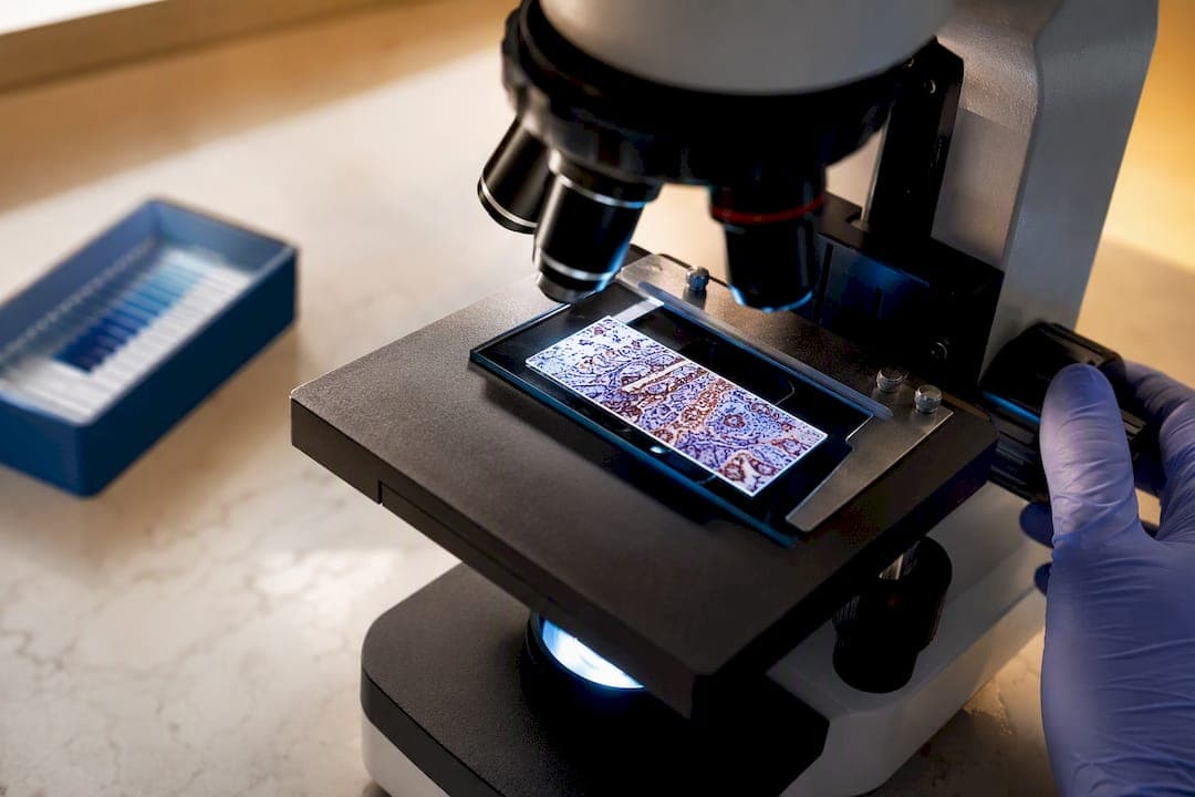
Immunohistochemistry is a laboratory technique that uses antibodies to detect and locate specific proteins inside tissue samples, letting pathologists see exactly which cells express a marker and where. Its main clinical job is diagnostic: it identifies what type of tumor a patient has, where it likely started, and sometimes which treatments will work. The sections below walk through how the test is actually run and how a pathologist reads the results.
TL;DR:
- Most IHC markers need standardized fixation times, with over- or under-fixation risking false negatives or weak staining results.
- Validation involves using known positive and negative tissues, with controls essential to confirm antibody specificity and detection accuracy.
- The type of antibody clone (monoclonal or polyclonal) directly impacts test specificity and sensitivity, requiring careful selection and validation.
- Automated platforms improve consistency, but manual optimization is still necessary for new or unusual markers before routine clinical use.
- Turnaround time varies depending on panel complexity, with single-marker tests generally faster than multi-marker or research-oriented stains.
Table of Contents
- What Is Immunohistochemistry, in Plain Terms?
- When Do Clinicians and Researchers Actually Order IHC?
- How Does a Tissue Sample Become a Stained Slide?
- Antibodies, Amplification, and Where IHC Goes Wrong
- How Do Pathologists Read an IHC Stain?
- How Labs Protect the Accuracy of an IHC Result
- What Patients and Trainees Should Actually Ask
- Getting IHC Testing Done Through EIV Diagnostics
- Authoritative Reading and Primary Sources
- Sources
- FAQ
What Is Immunohistochemistry, in Plain Terms?
Break the word apart and it explains itself. “Immuno” refers to antibodies, the immune system’s own targeting molecules. “Histo” means tissue. “Chemistry” is the reaction that makes an invisible antibody binding event visible under a microscope. Put together, immunohistochemistry (IHC) is a way of tagging specific proteins inside a tissue slice with antibodies, then triggering a color reaction so a pathologist can see exactly where those proteins sit.
Think of it like a highlighter that only marks one specific word on a page, no matter how many other words look similar. A standard hematoxylin and eosin (H&E) stain, the first stain done on almost every biopsy, shows overall tissue structure and cell shape in shades of pink and purple. It’s excellent for spotting abnormal architecture but blind to molecular identity. IHC picks up where H&E leaves off, answering the question H&E can’t: what protein is this cell actually making?
That distinction matters because two tumors can look nearly identical under H&E and still be biologically unrelated. Molecular tests like PCR or next-generation sequencing go a step further, reading DNA or RNA sequences directly, but they typically require the tissue to be dissolved or extracted, which destroys the architecture. IHC keeps the tissue intact while still answering a protein-level question, which is why it remains the workhorse test between basic morphology and full molecular profiling.
The technique itself isn’t new. Early versions used fluorescent tags in the 1940s, but fluorescence required specialized microscopes and faded quickly. The shift to chromogenic detection (permanent color stains visible on a standard light microscope) is what made IHC practical for routine diagnostic pathology, and it’s still the dominant approach in most labs today. A few things distinguish IHC from other pathology methods:
- It preserves the tissue’s cellular architecture, so location matters as much as presence or absence.
- It uses antibodies as the detection tool, not dyes that bind indiscriminately to cell structures.
- It can be read on a standard light microscope once a chromogen like DAB is applied, no special equipment required.
- It sits between basic histology and molecular diagnostics, adding protein-level specificity without destroying the sample.
When Do Clinicians and Researchers Actually Order IHC?
The single biggest use of IHC is sorting out what a tumor actually is once H&E shows something abnormal but not definitive. A poorly differentiated tumor can look like a generic ball of atypical cells under standard staining. IHC panels distinguish a carcinoma from a lymphoma from a melanoma, and can often pinpoint which organ a metastatic tumor originated from when the primary site is unknown. This diagnostic role, along with prognosis and treatment-response prediction, is central across organ systems including lung, prostate, colon, skin, and the central nervous system.
IHC also catches things conventional staining misses entirely. It can flag small clusters of metastatic tumor cells hiding in lymph nodes or bone marrow that would slip past a routine H&E scan, which matters directly for staging decisions and surgical planning.
Beyond diagnosis, IHC drives treatment selection. A few marker categories illustrate the range:
- Hormone receptors (estrogen receptor, progesterone receptor) determine whether a breast cancer is likely to respond to hormone-blocking therapy.
- HER2 status decides whether a patient qualifies for HER2-targeted drugs, and it’s one of a handful of markers with a standardized, treatment-linked scoring system.
- Proliferation markers like Ki-67 estimate how fast a tumor is growing, feeding into prognosis and grading decisions.
- Immune checkpoint markers such as PD-L1 help predict response to immunotherapy in several cancer types.
Research applications lean on the same underlying chemistry but for different goals. Labs validating a new biomarker use IHC to confirm a protein is actually present in the tissue type they’re studying before building a larger clinical study around it. Co-localization studies use double staining, applying two antibodies with two different chromogens or fluorescent tags on the same slide, to see whether two proteins sit in the same cells or the same subcellular compartment. That’s a common step in cancer biology research trying to map signaling pathways.
One principle runs through nearly all of this: IHC works best as a targeted answer to a specific question, not a blanket screen. Pathologists build a problem-oriented antibody panel matched to the differential diagnosis in front of them, rather than running every available marker and hoping something turns up.
How Does a Tissue Sample Become a Stained Slide?
The path from biopsy to a readable slide runs through a fairly rigid sequence, and small deviations at almost any step can change the result. Here’s the practical walkthrough.
- Collection and ischemic time. The clock starts the moment blood supply to the tissue is cut off. The longer a sample sits before fixation, the more proteins degrade, which is why minimizing the delay between excision and fixation matters more than most people realize.
- Fixation. Tissue goes into 10% neutral buffered formalin, generally for around 24 hours at room temperature, with an adequate ratio of fixative volume to tissue size. This step locks proteins in place and stops enzymatic breakdown, but it also chemically masks the very antigens IHC needs to detect later.
- Embedding. Fixed tissue is dehydrated and embedded in paraffin wax, creating a solid block that can be sliced thinly and uniformly.
- Sectioning. A microtome cuts the block into sections roughly 4 micrometers thick, mounted onto glass slides.
- Antigen retrieval. Because formalin fixation masks antigens, the slide needs heat-induced epitope retrieval (autoclave, microwave, pressure cooker, or water bath) or, for select antigens, enzymatic digestion to re-expose the target protein. The right method and timing has to be worked out empirically for each antigen-antibody pair, since there’s no universal setting that works for every marker.
- Blocking. A blocking reagent is applied to reduce background staining from nonspecific antibody binding, which would otherwise muddy the interpretation.
- Primary antibody incubation. The antibody targeting the specific protein of interest is applied and allowed to bind.
- Secondary antibody and detection. A secondary antibody, often linked to an enzyme system, binds the primary antibody, and a chromogen like DAB (producing a brown signal) or an AP red chromogen reveals the reaction site.
- Counterstain. A light hematoxylin counterstain colors the nuclei of all cells, giving the pathologist tissue context around the specific stain.
- Coverslipping and review. The slide is sealed and handed to the pathologist for interpretation under the microscope.
Pro Tip: If a report mentions “antigen retrieval failure” or unexpectedly weak staining, ask whether the tissue’s fixation time was documented. Under- or over-fixed tissue is one of the most common, and most preventable, causes of an inconclusive IHC result.
Manual staining protocols commonly run primary and secondary antibody incubations for 30 to 60 minutes at room temperature, though automated platforms in high-volume labs often compress or standardize these times differently. Every stage in this sequence also needs a control tissue run in parallel, a known positive sample confirming the antibody and detection system are working, and a known negative confirming there’s no nonspecific background staining. Without both, a stained slide tells you nothing reliable.
Pre-analytical variables deserve particular attention because they happen before the lab even sees the tissue. Variation in ischemic time and fixation duration measurably alters staining results for markers like estrogen receptor, progesterone receptor, HER2, and Ki-67, which is exactly why standardized fixation protocols exist for these clinically actionable markers in particular.
Antibodies, Amplification, and Where IHC Goes Wrong
Not all antibodies are built the same way, and the difference affects both specificity and how a result should be trusted. Monoclonal antibodies come from a single immune cell line and bind one specific epitope, giving high specificity but sometimes narrower reactivity across tissue variants. Polyclonal antibodies are a mixture of antibodies targeting several epitopes on the same protein, offering more sensitivity but a higher chance of cross-reactivity. This is why the specific clone name on a pathology report (something like “clone SP263” or “clone 3C6”) isn’t just paperwork. Different clones targeting the same protein can behave differently, and a lab switching clones without revalidating can shift results without anyone noticing until a discrepancy shows up.
Detection sensitivity also depends on amplification chemistry. Polymer-based detection systems link multiple enzyme molecules to a single secondary antibody, boosting signal without adding much background noise. Tyramine-based amplification pushes sensitivity even further for low-abundance targets, though it requires more careful optimization to avoid overamplifying background.
Errors creep in from both directions:
- False positives often come from endogenous peroxidase activity (common in tissues rich in red blood cells), incomplete blocking, or antibody cross-reactivity with an unrelated but structurally similar protein.
- False negatives frequently trace back to inadequate antigen retrieval, degraded antigens from prolonged fixation or delayed processing, or an antibody concentration that’s too dilute for the tissue type being tested.
- Uneven staining across a single slide can signal inconsistent fixative penetration, especially in larger or fattier specimens.
Validating a new antibody before it goes into clinical use means running it against known positive and negative tissues, confirming the staining pattern matches published literature, and titrating the concentration to find the point where specific signal is strong and background is minimal. A quick troubleshooting checklist for a suspect result: confirm the positive control worked, check the negative control for background, verify fixation time against the lab’s protocol, and confirm the antibody clone and lot number match what was validated. Manual optimization still matters here. Automated platforms improve consistency for routine panels, but newly introduced or unusual markers often need hands-on adjustment before they’re reliable enough for a clinical panel.
How Do Pathologists Read an IHC Stain?
Where the stain shows up inside the cell is often as diagnostically important as whether it shows up at all. A nuclear pattern (common for hormone receptors and proliferation markers like Ki-67) points to proteins involved in gene regulation or cell division. A membranous pattern (typical of HER2 and many immune markers) reflects proteins sitting on the cell surface, often receptors. A cytoplasmic pattern shows up for structural or metabolic proteins distributed through the cell body. A marker expressed in the wrong compartment for its expected biology is itself a red flag worth double-checking. This architectural context is exactly what IHC preserves and other molecular tests typically discard.
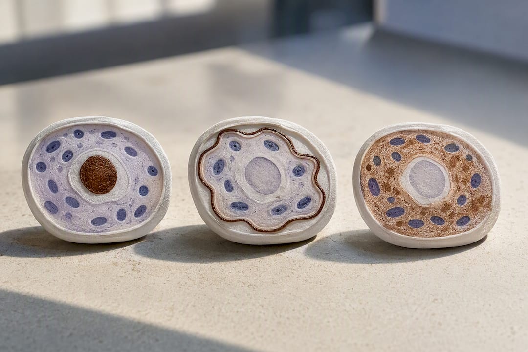
Reporting goes beyond a simple positive or negative call. Pathologists typically note staining intensity (weak, moderate, strong), the proportion of cells stained, and for some markers, a formal scoring system. HER2 and hormone receptor testing in breast cancer use standardized, validated scoring criteria directly tied to treatment eligibility, which is part of why labs can’t casually improvise a scoring approach for those particular markers.
Common marker categories and what they typically distinguish:
- Epithelial markers (cytokeratins) confirm a tumor arose from epithelial tissue, pointing toward a carcinoma.
- Melanocytic markers (S100, Melan-A) identify melanoma when a tumor’s origin is ambiguous under H&E.
- Lymphoid markers (CD20, CD3) separate B-cell from T-cell lymphomas and rule carcinomas in or out.
- Hormone receptor markers guide breast cancer treatment planning.
- Proliferation markers like Ki-67 estimate tumor growth rate for grading.
A positive result means the targeted protein is present in the tested tissue at a detectable level, not that a diagnosis is automatically confirmed. IHC results are meant to be interpreted alongside tissue morphology, clinical history, and other tests, functioning as a confirmatory step after H&E rather than a standalone verdict.
How Labs Protect the Accuracy of an IHC Result
Automation has changed how consistently IHC gets performed across a busy clinical caseload. Automated staining platforms standardize reagent volumes, incubation times, and washing steps, cutting down the variability that comes from technologist to technologist differences. That consistency matters most for high-volume, well-established panels. Manual staining still earns its place for research applications and for newly validated antibodies that need hands-on adjustment before they’re ready for routine use, since flexibility in optimization is harder to build into a fixed automated protocol.
Accreditation and internal validation are what keep a new marker from reaching a patient report before it’s ready. Every new antibody needs its own validation study against known controls, and lab accreditation standards exist precisely to audit whether that validation actually happened and is documented. Board-certified pathologists reviewing each case add a layer of clinical judgment that automated staining alone can’t replace.
Turnaround time is worth asking about directly. Clinicians and patients should ask a lab what its typical turnaround is for the specific panel ordered, since a straightforward single-marker stain moves faster than a complex multi-marker workup requiring digital slide review and pathologist consultation.
What Patients and Trainees Should Actually Ask
If a pathology report mentions IHC, ask your clinician which specific markers were tested and what a positive or negative result changes about your treatment plan. Don’t assume “positive” means worse news. It often just means a targeted therapy is now on the table.
If you’re learning the technique, pay closer attention to controls and fixation than to the chromogen step. Most staining problems trace back to what happened before the antibody ever touched the slide. Push your attending or mentor to explain the reasoning behind panel choices, not just the protocol steps. Good pathology depends on clinicians and pathologists talking through the clinical question together, not just exchanging a report.
— EIV Diagnostics
Getting IHC Testing Done Through EIV Diagnostics
If a biopsy report has left you or your care team needing a clearer answer, EIV Diagnostics runs the diagnostic services that IHC results usually feed into next. EIV Diagnostics offers histopathology for tissue-level diagnosis, molecular pathology when a case calls for DNA or RNA level answers alongside protein markers, and digital pathology for cases that benefit from remote specialist review. For skin-related findings, dermatopathology services are also available.

Whether you’re a provider ordering on behalf of a patient or someone self-paying for direct testing, have the specimen type, the specific clinical question, and any prior pathology reports ready before you request testing. That context helps the lab select the right antibody panel instead of running a broad, unfocused screen. The lab uses board-certified pathologists for interpretation and performs internal validation before new markers reach a patient report. Convenient mobile phlebotomy services allow specimen or blood collection to occur at home or in an office instead of requiring a separate lab visit. Explore the full range of services, including confocal microscopy and cytology, or check self-pay testing options to start the process directly.
Authoritative Reading and Primary Sources
For deeper technical detail, see the peer-reviewed protocol review Immunohistochemistry for Pathologists, the clinical overview on Immunohistochemistry applications, the antigen retrieval reference An Introduction to the Performance of Immunohistochemistry, and the patient-facing explainer from Cleveland Clinic.
This article is general information, not a substitute for advice from a qualified doctor. Consult a qualified healthcare professional about your own circumstances before acting on anything here.
Sources
FAQ
What Does IHC Reveal After a Biopsy?
IHC shows whether specific proteins are present in the biopsied tissue and exactly where inside the cells they’re located, helping pathologists classify the tumor type, its likely origin, and sometimes which treatments are appropriate.
What Does It Mean if an Immunohistochemistry Test Is Positive?
A positive result means the targeted protein was detected in the tissue. It doesn’t automatically confirm a diagnosis on its own. Pathologists interpret it alongside tissue architecture, clinical history, and other test results.
What Diseases Can Be Diagnosed by Immunohistochemistry?
IHC is used most heavily in cancer diagnosis, helping classify carcinomas, lymphomas, melanomas, and other tumors across organ systems including breast, lung, prostate, colon, and skin, and it also helps detect metastatic cells missed by standard staining.
How Long Do Immunohistochemistry Results Take?
Turnaround varies by panel complexity. A single-marker stain typically moves faster than a multi-marker workup requiring extended pathologist review. Patients and clinicians should check with their chosen testing lab for expected turnaround times on specific panels.
How Is Immunohistochemistry Different From Immunofluorescence?
Both use antibodies to detect specific proteins, but IHC typically uses a chromogen like DAB for a permanent color visible under a standard light microscope, while immunofluorescence uses fluorescent tags that require a fluorescence microscope and tend to fade over time.